Download
1 / 60
620 likes | 975 Views
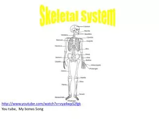
Skeletal System. http://www.youtube.com/watch?v=vya4wpS2fgk You tube, My bones Song. Chapter 5: The Skeletal System. I : Bones: An Overview A. There are 206 bones in the human body, which makes up about 20% of our body mass. B. The parts of the skeletal system include:

E N D
Skeletal System http://www.youtube.com/watch?v=vya4wpS2fgk You tube, My bones Song
Chapter 5: The Skeletal System I: Bones: An Overview A. There are 206 bones in the human body, which makes up about 20% of our body mass. B. The parts of the skeletal system include: 1. Bones- internal framework that supports and anchors all organs. 2. Joints- allow for flexibility and movement 3. Cartilages- provides additional support for structures in the body. 4. Ligaments- connect bone to bone. C. The skeletal system is subdivided into two groups: Axial and Appendicular. 1. Axial Skeleton- includes the skull, vertebral column and rib cage. 2. Appendicular Skeleton- includes bone of the upper and lower limbs.
D: Functions of the Bones 1. Support- bones act as pillars to support the body 2. Protection- for soft body organs 3. Storage- for fat in the internal cavities (yellow marrow), also bones store minerals such as calcium and phosphorous 4. Blood Cell Formation (Hematopoiesis) - blood cells are formed in the red marrow of certain bones (Newborns contain only red marrows. in adults about half of the marrow is red and located in -ribs, breastbone, shoulder blades, collarbones, hip bones, skull, and spine..
E: Two Types of Tissues 1. Compact bone- the external layer that is dense and looks smooth and homogeneous a. Contains the functioning units of bone called osteons. 2. Spongy bone- the internal layer that is composed of a honeycomb of needle like or flat pieces called trabeculae. a. The open spaces between trabeculae are filled with red or yellow bone marrow
F: Chemical Composition of Bone- inorganic and organic components 1. Inorganic- 65% of bone mass consists of hydroxyapatites or mineral salts (largely calcium phosphates) a. Calcium salts account for the exceptional hardness of bone and allows it to resist compression. Foods high in calcium: Foods high in phosphorous: Foods that are high in phosphorus include milk (234mg per 8 ounces), milk products, poultry, fish, meat, eggs, grains and legumes.
2. Organic- cells, osteoblasts (bone making cells), and osteoid. a. Osteoid makes up 33% of bone matrix and includes proteoglycans, glycoproteins and collagen fibers. Major component of extracellular matrix b. Organic components allow for bone flexibility and great tensile strength to allow bone to resist stretch and twisting motions.
G. Classification of bones according to shape 1. Long Bones a. Typically longer than wide b. Have a shaft with heads at both ends c. Contain mostly compact bone d. Examples: femur and humerus 2. Short Bones a. Generally cube shaped b. Contain mostly spongy bone c. Examples: carpals and tarsals d. Sesamoid bones are special type of short bones that are embedded in a tendon. Ex. Patella
3. Flat Bones a. Thin and flattened b. Usually curved c. Thin layers of compact bone around spongy bone d. Example: skull, scapular, ribs. Sternum 4. Irregular Bones a. Irregular shape that does not fit other bone classifications b. Example: vertebrae , sacrum, mandible
Mr. Bones Apart- pg. 5M • Articulate Mr. Bones (cut out his bones and put him back together. All bones should be connected.) • Color his axial skeleton Red and his appendicular skeleton Blue • Label two: long bones, short bones, irregular, and flat bones • Label the bones with their name. • Write the functions of the skeletal system around Mr. Bones. • Use the cards on pgs. 5I and J to write the meaning of the bone name • Answer questions on pg.5L
Question of the Week • Why is it that once we sprain our ankle we are more likely to sprain the same ankle again? The ligaments in your ankle, are laced with sensory receptors. These are responsible for telling the brain where the ankle is in space. When a sprain occurs, some of these sensors are permanently damaged; as a result, your ankle can't communicate as well with your brain. Rehab, when approached correctly, can help you regain a kind of ankle virginity--that is, if you remain sprain-free for a year, your risk returns to what it was before your mishap.
II. Bone Structure A. Gross Anatomy of a typical long bone 1. Diaphysis - the bone shaft that makes up most of the bones length and is composed of compact bone a. The diaphysis is covered with a fibrous connective tissue membrane called the periosteum. Sharpey’s Fibers (connectivetissues) secure the periosteum to long bone. b. The diaphysis surrounds a central medullary cavity or marrow cavity (yellow marrow) primarily used for storing adipose (fat) tissue. In infants this area forms blood cells, and red marrow is found here. In adults, the red marrow is confined to the cavitiesof spongy bone of flat bones and the epiphyses of long bones
2. Epiphysis- the ends of long bones a. Consists of a thin layer of compact bone enclosing an area filled with spongy bone b. In adult bones, a thin line of bony tissue spans the epiphysis and separates it from the rest of the bone in that area, this is called the epiphyseal line. The epiphyseal line is a remnant of the epiphyseal plate (flat plate of hyaline cartilage) you would see in a young growing bone.
Anatomy of a Long Bone spongy bone Proximal epiphysis compact bone Endosteum diaphysis epiphyseal line yellow marrow Sharpey’s fibers Distal epiphysis hyaline cartilage periosteum
3. Bone Markings the bumps, holes, and ridges seen on bones and reveal where muscles, ligaments, and tendons attach. There are two categories of bone markings: a. Projections or Processes - grow out from the bone surface, (see chart on pg.119) Ex. Spinous process of vertebrae b. Depressions or cavities - indentions in the bone, all depressions begin with the letter F; ex. Foramen magnum of the base of the skull
B. Microscopic Anatomy of Compact Bone 1. Osteocytes - Mature bone cells 2. Osteons - units of bone (Haversian System) 3. Haversian CANAL a. opening in the center of an osteon b. Run lengthwise through the bone carrying blood vessels and nerves to all areas of the bone 4. Perforating (Volkmann’s) canals a. perpendicular to central canals b. carries blood vessels and nerves http://www.youtube.com/watch?v=4qTiw8lyYbs&feature=related
5. Lacunae - tiny cavities a. contain bone cells (osteocytes) b. arranged in concentric rings 6. Lamellae - rings around the central canal a. sites of lacunae 7. Canaliculi - tiny canals that radiate from central canals to connect all bone cells to the nutrient supply
Microslide of Bone Tissue • Get a microslide viewer to share with someone at your lab station. • Draw and label slide 4 of bone tissue. Label this as MICROSCOPIC bone anatomy. This picture should take up half of a sheet of notebook paper. Make sure it is neat and labeled using the specific names we discussed in the notes today. • Answer the following questions on your paper • What are the chemicals found in our cells that make them strong? • What are the fine dark lines connecting each of the bone cells? • The dark area labeled B is used for what purpose? Label the specific name for this structure. • Explain what happens to the bones of someone with osteoporosis. • What do you think would look different about the bone tissue in a patient with osteoporosis. Explain or draw a picture (use words we have learned today )
Choose a long bone in the body and complete oneof the choices below: 40 pts_____ Write an advertisement (as if the bone is describing and selling itself and its importance)_____ Story from the bones perspective (The Bony Life of Henrietta Humerus, an Autobiography) • _____ Facebook Profile Page (Ex. Freddy Femur)_____ Create a flow chart/ Concept Map with pictures and diagrams • Regardless of which you choose YOUR FINAL PRODUCT MUST: • Explain the structure and function of the following parts of the bone you choose: • diaphysis, epiphysis, sharpey’s fibers, periosteum, cartilage, osteon, osteocytes, osteoblasts, osteoclasts • 20pts. (1pt. For structure, one for function) • What is your chemical composition and their functions (organic and inorganic)? 4pts. • How do you fit into the entire skeleton? What bones do you articulate (come together) with? 6 pts. • What movements are allowed by this bone and the bones it articulates with? 5 pts. • What muscles attach to your bone? 5 pts.
Microscopic Bone Anatomy C- H- L-
III. Bone Formation and Growth A. Bone formation is also known as osteogenesis (bone beginning) ossification. 1. Formation of the skeleton a. In embryos the skeleton is composed mainly hyaline cartilage (fibrous membranes at skull and clavicles) b. Intramembranous ossification process in which the bones form toreplace fibrous membranes of the skull and clavicles.
275 bones12 weeks (6-9 inches long) Fetal Skeleton
Fetal Skull Intramembranous Ossification - Fontanels (membranes) become bone.
Fetus: 1st 2 months Endochondral Ossification = Cartilage to bone 2o ossification center bone cartilage calcified cartilage Just before birth epiphyseal line epiphyseal plate Childhood Adult
C. Endochondral ossification- process in which hyaline cartilage is replaced by bone (includes all bones except skull and clavicles) i. The hyaline cartilage is covered in osteoblasts (bone formingcells) ii. The Fetus then has cartilage “bones” enclosed by bony bones iii. The enclosed hyaline cartilage is digested away, opening up a medullary cavity within the newly formed bone. 2. Cartilage remains in the bridge of the nose, parts of the ribs, joints (articular cartilage), and epiphyseal plates. http://wps.aw.com/bc_martini_eap_4/40/10466/2679495.cw/content/index.html
Chinese Binding of Feethttp://www.historyforkids.org/learn/china/clothing/How do you think binding of the feet affected bone growth?
B. Bone growth- occurs at epiphyseal plates 1. Provides for longitudinal bone growth during childhood 2. Controlled by growth hormones and sex hormones 3. New Cartilage is continuously formed 4. Old cartilage becomes ossified a. Cartilage is broken down and replaced by bone
Differences in fetal skeleton vs. Adult 1. Abundance of cartilage vs. bone 2. Incomplete or lack of fusion between bones 3. Poor development based on lack of use 4. Large head size compare to the rest of body ( principle of cephalization) 5. Facial vs. Cranial parts of the skull 6. Frontal bone and the metopic suture 7. Fontanels 8. Pelvic bone 9. Spinal curvatures On average, an adult human has 206 bones (according to Gray's Anatomy, but the number can vary slightly from individual to individual), but a baby is born with approximately 270 -275 bones.
5. Growing bones must also widen as they lengthen (called appositional growth) a. osteoblasts begin adding bone to the outside of the diaphysis b. osteoclasts (bone destroying cells) in the endosteum remove bone from the inside diaphysis wall c. The work of osteoblasts and osteoclasts occur at almost the same rate allowing the bone to expand and widen
Body Ratios and Proportions (pg. 5U) and write a one page summary including the following: • Explain the changes to our skeletal system throughout our lifetime (osteogenesis to bone growth to changes in old age). Include data from your measurements to determine if you are still growing and how you used the data to come to this conclusion.
Bone Anatomy Quiz Osteoctyes Canaliculi Lacunae Haversian Canal Osteon (Haversian system) 1. The entire structure pictured (functioning unit of bone) is called? 2. identify H 3. identify L 4. identify C
Epiphysis, diaphysis, articular cartilage, spongy bone, compact bone D. Structure A C. Type of Bone? . Structure B D 5. Identify A 6. Identify B 7. C- what type of bone? 8. Identify D Organic or Inorganic Bone components? 9. Provides hardness 10. Allows for flexibility and tensile strength
11. Growth in bones occurs at _________plates. • Function of red marrow? • Function of yellow marrow? • Which term does not belong? Lamellae----- canaliculi-----circulation----osteoblasts
How much calcium is in that? • http://www.fitsugar.com/How-Much-Calcium-182622
Bone cells that aid in remodeling Builds new bone Osteoblast Mature bone cell Osteocyte Osteoclast Eats bone
IV. Bone Remodeling- the two processes of bone deposits and removal A. In an adult skeleton bone deposits and bone removal are occurring all the time (yes, bone is a dynamic, active living tissue) B. In a healthy adult the rate of bone deposit and bone removal should remain constant and equal 1. Bone deposits occur where bone is injured or additional bone strength isneeded a. Deposits are accomplished through osteoblasts b. Optimal bone deposits require a diet rich in proteins, vitamin C, A, and B12, calcium, phosphorus, and magnesium 20% of your skeleton is replaced annually!! Entire skeleton is replaced every 7 to 10 years!
2. Bone Removal or resorption occurs when the blood levels of calcium become too low. a. Resorption is accomplished by osteoclasts 3. If bone removal (resorption) occurs faster than bone deposition takes place homeostatic balance is lost. This results in porous, lightweight bones that easily break. This condition is called osteoporosis. This occurs most often in aged, postmenopausal women, but can occur in both sexes.
C. Bone remodeling is controlled by hormones, diet and mechanical stress 1. Hormonal mechanism- involves the parathyroid hormone (PTH) and calcitonin in a negative feedback loop to maintain homeostasis of blood calcium.
a. PTH is released when blood calcium levels are too low. It stimulates osteoclasts to resorb (remove) bone releasing the calcium into the blood. b. When blood calcium levels rise calcitonin is secreted to encourage calcium deposits into the bone. 2. Diet a. Calcium is necessary for transmission of nerve impulses, muscle contraction,blood coagulation, secretion by gland and nerve cells, and cell division i. Your daily recommend levels of calcium is 1200-1400 mg. ii. Calcium is absorbed by the intestines under the control of vitamin D metabolites 3. Mechanical Stress a. This set of controls serves the needs of the skeleton itself- keeping bones strong where stressors are present. b. Wolff’s Law - holds that bones grow or remodel in response to the forces or demands placed on it. Bone is laid down where bone is needed- for example large bony projections occur where heavy, active muscles attach
Steps in Bone RemodelingQuiescence means inactive, or dormant
Flashcard Warm-up March 8th #30 Bone Remodeling Three factors that affect bone remodeling are ____, _____ _____, and _____. Steps in the process are: (refer to your bone remodeling and briefly explain)
V. Bone Fractures A. A break in the bone. There are two types: 1. Open (compound) fracture - the broken bone penetrates through the skin 2.Closed (simple) fracture - the broken bone does not penetrate through the skin B. Bone fractures are treated by reduction and immobilization 1. Realignment of the bone. C. Common types - comminuted , compression, depression, compacted, spiral and green stick - see book for pictures
Repair of Fractures hematoma callus bony callus bone remodeling
D. Repair of Bone Structures 1. After the fracture occurs, blood vessels rupture forming a blood - filledswelling called a hematoma. 2. A fibrocartilage callus forms (made of various connective tissues) and acts to splint the broken bone 3. A bony callus forms as osteoblasts replace the fibrocartilage with bone. 4. The bony callus continues to be remodeled in response to mechanical stress placed upon the bone.
I. E. Repair of Breaks • 6-8 hr.: hematoma develops, blood to site of break, Leukocytes (WBC, fight infection, swelling


