Download

1 / 19
190 likes | 412 Views
Retina Conference. Janelle Fassbender , MD, PhD University of Louisville Department of Ophthalmology and Visual Sciences 01/23/2014. Subjective. CC/HPI : 52 year old HF c/o the right half of faces looking abnormal. POH : None PMH : Hypertension, hyperlipidemia, type 2 DM

E N D
Retina Conference Janelle Fassbender, MD, PhD University of Louisville Department of Ophthalmology and Visual Sciences 01/23/2014
Subjective CC/HPI: 52 year old HF c/o the right half of faces looking abnormal. POH: None PMH: Hypertension, hyperlipidemia, type 2 DM Meds: Metformin, 3 anti-hypertensive agents, omeprazole, prevastatin FOH: Mother with macular hole
Objective OD OS Va(sc): 20/20-2 20/50-2 Pupils: 3 No RAPD 3 IOP: 20 19 EOM: Full Full
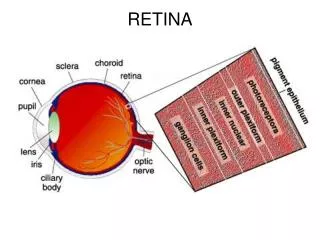
Anterior and Posterior Segments OD OS Anterior segment: WNL OU ON: c/d 0.2 pink/sharp c/d 0.2 pink/sharp Macula: Cystic foveal lesion Cystic foveal lesion Vessels: WNL WNL Periphery:WNL WNL
OCT OD OS
OCT OD Parafoveal intra-retinal cavity extending to and involving the outer retina; irregularity of the ELM, focal discontinuity of the photoreceptor ellipsoid line and cone outer segment sheaths
OCT OS Intra-retinal cystic foveal lesion extending to RPE with interruption of the photoreceptor outer segments.
Fluorescein angiogram • OD: Mid A-V phase with leakage temporal to fovea.
Fluorescein angiogram • OS: Temporal leakage and focal hyperfluorescence.
Differential Diagnosis • Parafovealtelangiectasia • Non-Proliferative Diabetic Retinopathy • Stage 1 macular hole • Hypertensive retinopathy • Branch retinal vein occlusion
Diagnosis Parafovealtelangiectasia
Plan Follow up in 4-5 months.
Parafoveal telangiectasia • Also known as idiopathic juxtafoveolar retinal telangiectasisor idiopathic macular telangiectasia • First described by Gassin 1968 • Initial classification in 1982 by Gass and Oyakawa • Heterogeneous group of disorders classified into 3 types with independent etiologies. • Prevalence of 0.1% per Beaver Dam Eye Study (2010)
Pathophysiology • No actual telangiectasis – vessel wall thickening with eventual capillary dilatation (Green et al, 1980; Gasset al, 1982). • Metabolic alteration, endothelial permeability, nutritional deficiency degeneration of the middle and outer retina. • Neural, Muller or endothelial cell etiology (Cohen etal,2007)? • Crystalline deposit – degenerated Muller footplates • Fluorescein leakage – loss of Muller-mediated barrier • Outer retinal atrophy – loss of Muller cell nutritional and mechanical support • Cystoid spaces of type 1 compared to 2, retinal vein occlusion, and diabetic macular edema (Oh et al, ePub ahead).
MacTel Project (2005) • International consortium to study cause, natural history, progression and epidemiology • Resulted in multiple publications describing clinical findings, diagnostic methods, and epidemiology. • 27 candidate genes – None associated to MacTel(Parmalee et al, 2010). • Genome-wide linkage study (Parmalee et al, 2012): • Probable AD transmission with reduced penetrance and expressivity. • Linked to 1q41-42 (LOD 3.45)
Pathophysiology • ATM was characterized in 1988 as the causal gene for ataxia telangiectasia (AT) • Chronic oxidative stress resulting in DNA damage activates ATM and leads to increased apoptotic activity. • Loss of ATM function leads to genome instability • Allelic variants noted in 13/30 macular telangiectasia, especially of European ancestry (Barbezetto, 2008). • 11/16 with polypoidalchoroidalvasculopathy or macular telangiectasia (Mauget-Faysee, 2003)
Bevacizumab therapy for idiopathic macular telangiectasia type IIKovach and Rosenfeld, Retina. 2009 Jan;29(1):27-32 • Purpose: To determine if inhibition of VEGF-A affects visual acuity, fluorescein angiographic (FA), and optical coherence tomography (OCT) outcomes in patients with perifoveal telangiectasia (PT) type 2A. • Results: 9 eyes of 8 patients. After treatment, follow-up ranged from 4 to 27 months. • Non-proliferative - Mean BCVA remained stable (n = 4). • Proliferative - BCVA was unchanged or improved after treatment (n = 5). • All eyes demonstrated decreased intraretinal leakage on FA after an injection of bevacizumab, and eyes with proliferative PT showed decreased growth and leakage of the subretinal neovascularization. • The mean decrease in OCT central retinal thickness – 6 um non-proliferative; 26 um proliferative.
Conclusions: IntravitrealAvastin • Non-proliferative PT: • Decreases fluorescein angiographic leakage in PT but has no short-term effect on visual acuity or OCT appearance. • Proliferative PT: • Arrests the leakage and growth of subretinal neovascularization with the possibility of visual acuity improvement.
References • Oh, JH, et al. 2013. Characteristics of cystoid spaces in type 2 idiopathic macular telangiectasia on spectral domain OCT. Retina. [Epub ahead of print] • Wu, Evans, Arevalo. 2013. Idiopathic macular telangiectasia type 2. SurvOphthalmol, 58(6):536-59 • Mauget-Faysse, et al. 2003. Idiopathic and radiation-induced ocular telangiectasia: the involvement of the ATM gene. Invest Ophthalmol Vis Sci. 44(8):3257-62. • Barbazetto IA, et al. 2008. ATM gene variants in patients with idiopathic perifoveal telangiectasia. Invest Ophthalmol Vis Sci, 49(9):3806-11. • Kovach and Rosenfeld. 2009.Bevacizumab therapy for idiopathic macular telangiectasia type II. Retina. Jan;29(1):27-3. • Sawsan, et al. 2010. Idiopathic juxtafoveolar telangiectasia: A current review. Middle East Afr J Ophthalmol, 17(3). • Yannuzzi et al. 2006. Idiopathic macular telangiectasia. JAMA Ophthalmol, 124(4):450-460. • Parmalee, et al. 2010. Analysis of candidate genes for macular telangiectasia type 2. Molecular Vision, 16:2718-26. • Parmalee, et al. 2012. Identification of a potential susceptibility locus for macular telangiectasia type 2. PLOS One, 7(8). • Cohen et al. 2007. Optical coherence tomography findings in nonproliferative group 2a juxtafoveal retinal telangiectasis. Retina, 27(1):59-66.