Download
1 / 32
330 likes | 480 Views
The retina is a crucial component of the eye, containing millions of light-sensitive cells, including rods and cones. Rods are highly sensitive to light and allow for vision in low-light conditions, while cones provide color vision and visual acuity in brighter settings. Rhodopsin in rods and iodopsin in cones play significant roles in generating action potentials in response to light. Additionally, the retina undergoes adaptations to adjust between light and dark conditions, enhancing our ability to perceive stimuli. This overview explores the structure, function, and differences between rods and cones.

E N D
The Retina • WALT • That the retina contains millions of light sensitive cells • That there are two types of light sensitive cell • How an action potential is generated
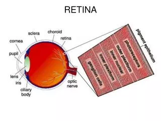
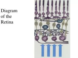
The retina • The retina is a layer at the back of the eye which is sensitive to light • It contains two types of light sensitive cells • The rods • The cones • They connect to bipolar neurone which connect to ganglion cells. • The fibres of the ganglion cells form the optic nerve
Structure of a single rod cell False colour scanning EM of cone and rod cells
Rhodopsin • Each rod possesses up to a thousand vesicles in its outer segment containing rhodopsin. • Rhodopsin is made up of the protein opsin and retinal (a derivative of vitamin A). • Retinal normally exists in its cis isomer form, but light causes it be become converted to its trans isomer form. • This change initiates reactions which lead to the splitting of rhodopsin into opsin and retinal - bleaching.
Action Potential Generation • The free opsin acts as an enzyme which sets of a series of reactions that leads to the hyperpolarisiation of the rod cell membrane • It becomes MORE NEGATIVE • This generates an action potential is the connecting neurone cells
Light and Dark Adaptation • Rhodopsin is very sensitive to light • In bright light it is broken down faster than it can be resynthesised • It is “bleached” for most of the time • We are light adapted • If we go into a dark room we can’t see much until the rhodopsin is resynthesised and we become dark adapted.
Convergence • Several rod cells connect with a single bipolar neurone. This increases the ability of the brain to detect a small amount of light • This is called convergence and it allow for weak stimuli to be amplified giving rods a greater sensitivity
Rods and Cones • Rods out number cones • There are about 120 million rod cells • There are about 6 million cone cells
Cones • Each cone is connected to its own bipolar cell • This means that the eye is able to distinguish between two or more separate stimuli • This is called visual acuity – the amount of detail we see
Cones • Vision in bright or moderate light is brought about by the functioning of the cone cells. • Cone cells provide vision in colour as well as black and white • They enable you to see in fine detail – visual acuity
Cones • Cone cells contain a pigment called iodopsin and retinal • There are three types of iodopsin but only one type is found in each cone cell • 10% red cones • 45% blue cones • 45% green cones • Each type of iodopsin absorbs most strongly in a particular part of the visual spectrum
Colour Vision - Colour vision is due to the presence of 3 kinds of cone cells detecting 3 kinds of primary colours (trichromatic theory): red, blue and green
The Trichromatic Theory • Very few people cannot distinguish colours at all • Most colour blind people have abnormal colour vision • Some males have inherited their in ablilty to distinguish between reds and greens • The genes for red and green iodopsin are found on the X chromosome
Generation of action potential in Cone Cells • A similar process occurs in cone cells except that the pigment is iodopsin. • It is less sensitive to light and so a greater intensity is required to cause its breakdown and so initiate a nerve impulse.
Acuity • Cone cells are found throughout the retina • They are far more concentrated at a point called the fovea • As each cone cell synapses with one bipolar cell this allow us to see in detail • The fovea is the area of greatest visual acuity
The Blind Spot • There is a point on the retina where there are no rods or cones • This is the blind spot- it is where the optic nerve leaves the retina