
RETINA
E N D
Presentation Transcript
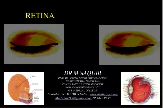
RETINA VIRAL KERATITIS DR M SAQUIB MBBS,MS , FSCEH DELHI,FHVDESAI PUNE, EX REGISTRARA JNMCH,AMU CONSULTANT OPHTHALMOLOGIST HOD D/O OPHTHALMOLOGY G.S .MEDICAL COLLEGE Founder sec: MEDICS India , www.medicsngo.org Mail-dms2k5@gmail.com , 9634123800
CONTENTS 1.ANATOMY 2.CONGENITAL & DEVELOPMENTAL DISORDER S 3.INFLAMMATORY DISORDER –Retinitis, Vasculitis 4.VASCULAR DISORDER OF RETINA – Vascular Occlusions Diabetic ,Hypertension ,sickle cell ,Prematurity Retinopathy , Retinopathy in Pregnancy ,Blood Disorders ,Retinal Telangiectasia, Ocular Ischemic Syndrome 5. RETINAL DYSTROPHIES & DEGENERATION 6.MACULAR DISORDER 7. RETINAL DETACHMENT – Rhegmetogenous ,Exudative, Tractional . 8.TUMOURS OF RETINA
Retina Developed from 2 walls of optic cupNervous Retina –From Inner wall Pigment Epithelium from Outer wall
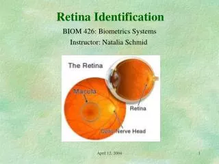
A. ANATOMY • Retina is a thin delicate transparent membrane forming the innermost covering of the eyeball and extending from the optic disc to the ora serrata. • Appears Purplish Red ( Rods& choroid) • Surface Area -266 mm2 • Thickness- Ora serrata -0.1mm Equator – 0.18 mm Peripapillary-0.56mm
MACULA LUTEA • 5.5 mm circular area deeper red than rest of the fundus at the posterior pole of retina,. • lying inside the temporal vascular arcades, 2 disc diameters temporal to the optic disc • Macula lutea (5.5mm) • Fovea centralis (1.5mm)- Most sensitive part of Retina ,Cones only • Foveola (0.35mm)- 2DD ( 3-4mm) from temporal margin og Disc and 1 mm below horizontal meridean • Umbo- Tiny depression . Shining foveal Reflex on examination. • Parafoveal region(o.5mm)- • Perifoveal region(1.5mm) • -Foveal Avascular Zone (FAZ)-0.8 mm Not contain any capillaries
Macula Lutea Fovea Centralis 1.5mm Parafovea 0.5mm wide Perifovea 1.5mm wide Foveola Diameter:0.35mm Umbo: Tiny Depression in the Very Centre of Foveola
Optic disc • Pale pink • Well defined circular area • 1.5 mm • Cup It varies in size, shape, position and depth in different eyes. • All the retinal layers terminate at the optic disc except the nerve fibres ,which pass through the lamina cribrosa to run into the optic nerve.
OPTIC DISC ( Optic Nerve Head ) • Oval area vertical-1.88 mm • Horizontal- 1.76 mm • Begining of Optic Nerve / Optic Nerve Head • Depression in Optic Disc – Physiological Cup • No Photoreceptors at Optic Disc – ABSOLUTE SCOTOMA PHYSIOLOGICAL BLINDSPOT • Nasal border is steeper & temporal border has a more gradual slope
Peripheral Retina • Area of Retina Ant –ora Serrata & Posteriorly by Equator . • Examined – I.O or Goldman Three Mirror contact Lens
Ora Serrata • Serrated Peripheral Margin • Anterior End of Retina • Retina Firm attachement with Vitreous & Choroid • Pars Plana Extends Anteriorly
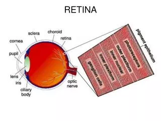
MICROSCOPIC STRUCTURE • 10 Layers • Pigment Epithelium • Neurosensory Retina – 3 types of cells Muller Amacarine Bipolar synapses
Retinal pigment epithelium(RPE) • Outermost Layer Of Retina • Provide metabolic support & Antireflective layer • Consisting Of A Single Layer Of Hexagonal Cells • Containing Pigment • Firmly Adherent To The Underlying Bruch’s Membrane of choroid • And Loosely Arranged To The Layer Of Rods And Cones • The Potential Space Between RPE And The Sensory Retina Is Called Subretinal Space • INTERPHOTORECEPTOR MATRIX (IPM) Present in the potential space binding support .
RPE Electron microscopy shows that adjacent RPE cells are connected with each other by tight junctions(zonulae occludens anzonulae adherens) and constitutes the outer blood-retinal barrier
PHOTORECEPTORS -Rods & ConesElectromagnetic wave of light to Electro chemical • 120 million rods • 6.5 million cones • Outer segment of photoreceptor cells Rods – 40-60 um,Scotopic Vision Visual Purple (Rhodopsin) Vision of low illumination Peripheral Vision Cones- Photopic Vision Colour Vision 40-80um long, longest at the fovea and shortest at the periphery.
External limiting membrane: • In low magnification, it appears as a fenestrated membrane extending from the ora serrata to the edge of the optic disc. • Electron microscopy studies show that ELM is formed by the junctions between the cell membranes of photoreceptors and mullers cell. • Process of Rods and Cons pass throgh
INNER NUCLEAR LAYER • This layer resembles the outer nuclear layer except it is very thin. It disappears at fovea and in rest of the retina consists of the following; • Bipolar cells,( First Order Neuron ) • Horizontal cells, • Amacrine cells, • soma of the Mullers cells, • Capillaries of central retinal vessels
BIPOLAR CELLS • These are the neurons of first order of vision.The body of the bipolar cells consists entirely of the nucleus which lies in the inner nuclear layer. Their dendrites arborize with the rod spherules and cone pedicles in the outer plexiform layer and their axons arborize with the dendrites of ganglion cells in the inner molecular layer.
MULLER CELLS • The nucleus and cell bodies of the mullers cells are located with in the inner nuclear layer.The mullers cell provide structural support and contribute to the metabolism of the sensory retina.
Inner Plexiform Layer This layer essentially consists of synapses between the axons of bipolar cells(first order neurons),dendrites of ganglion cells(second order neurons) and the processes of integrative amacrine cells. • This layer is absent at the foveola.
Ganglion cell layer • The cell bodies and the nuclei of the ganglion cells ( Second Order Neuron of visual pathway ) lie in this layer. • Monosynaptic Ganglion cells – • In macular region • Dendrites of each cell synapse with single Axon of Bipolar cells • Polysynaptic Ganglion Cells – • One dendrites may synapse with hundred of Axon cells . • Peripheral Retina
Nerve fibre layer • This layer consists of the unmyelinated axons of the ganglion cells which converge at the optic nerve head, pass through lamina cribrosa and become ensheathed by myelin posterior to lamina. • This layer also contains the follwing; • Centrifugal nerve fibres, • Processes of mullers cells, • The neuroglial cells present in the nerve fibre layer, • Retinal vessels lie in the nerve fibre layer.
Internal Limiting Membrane • It mainly consists of a PASpositive true basement membrane that forms the interface between retina and vitreous. It consists of four elements, Collagen fibrils, Proteoglycans(mostly hyaluronic acid)of the vitreous, • The basement membrane, • The plasma membrane of the mullers cells and possibly other glial cells of the retina.
Fovea Centralis • In this area, there are no rods, cones are larger,in abundance and tightly packed, and other layers of retina are very thin. • Its central part (foveola) largely consists of cones and their nuclei covered by a thin internal limiting membrane. • All other retinal layers are absent in foveolar region.
Henle’s layer – In the foveal region surrounding the foveola cone axons are arranged obliquely .
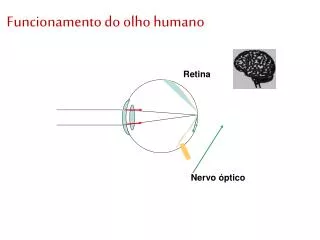
VASCULAR SUPPLY OF RETINA Outer four layers of the retina get their nutrition from the choriodal vessels. The six inner layers get their blood supply from the central retinal artery. The fovea is an avascular area mainly supplied by the choriocapillaries. The macular region gets its blood supply by small twigs from the superior and inferior temporal branches of central retinal artery. • The branches of central retinal artery are end arteries i.e they dont anastomose with each other.
Central Retinal Artery • Branch of ophthalmic Artery • Emerge from center of physiohlogical cup of disc • 4 brances – Sup –nasal,Su-Inf • Inf –Nasal-Inf Temp • End Artery
Retinal Veins • Follow pattern of Retinal Artery • Drains into cavernous sinus directly or through superior ophthalmic vein