THE RETINA
2.94k likes | 4.09k Views
THE RETINA. DR. AMER ISMAIL ABU IMARA JORDANIAN BOARD OF OPHTHALMOLOGY INTERNATIONAL COUNCILOF OPHTHALMOLOGY PALESTINIAN BOARD OF OPHTHALMOLOGY. Development of the retina The eye is externalized portion of the brain .

THE RETINA
E N D
Presentation Transcript
DR. AMER ISMAIL ABU IMARA JORDANIAN BOARD OF OPHTHALMOLOGY INTERNATIONAL COUNCILOF OPHTHALMOLOGY PALESTINIAN BOARD OF OPHTHALMOLOGY
Development of the retina • The eye is externalized portion of the brain . • Formation of the eye begins with lateral outpouchings of the forebrain during the third week of development . • The development of the optic cup ( optic vesicle ) reaches a stage where the outer layer of the optic vesicle becomes the retinal pigment epithelium , while the inner layer of the optic vesicle becomes the multilayered neurosensory retina . anterior extension of both layers become the double layer ciliary epithelium . • The ocular ventricle is the potential space between the retinal pigment epithelium and the neurosensory retina.
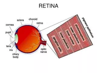
PIGMENT EPITHELIUM • Is homolog of the epithelium of the choroid plexus of the brain . • The retinal pigment epithelial cells acquire during development tight junctions that form a barrier between the neurosensory retina and the choriocapillaries .
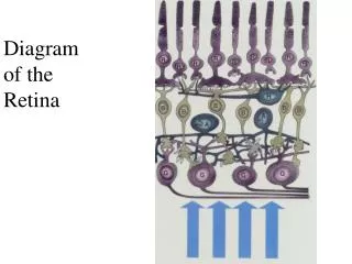
CELLULAR ORGANIZATION OF THE RETINA • The layers of cell nuclei are as follows : • the outer nuclear layer (ONL) , which contains the cell bodies of the photoreceptors. • The inner nuclear layer (INL) , which contains the cell bodies of horizontal neurons , bipolar neurons , amacrine neurons , displaced ganglion cells and those of the glial cells of Muller . • The ganglion cell layer , which contains the cell bodies of most of the ganglion cells , displaced amacrine cells and those of the astroglial cells .
Between the ONL and the INL is the outer plexiform layer (OPL) • OPL = synapses of the photoreceptors ,bipolar cells and horizontal cells . • Between the INL and the ganglion cell layer is the inner plexiform layer ( IPL) • IPL= synapses of the bipolar cells , amacrine cells and ganglion cells .
The optic fibers consist of the axons of the ganglion cells and are unmyelinated while within the retina . these fibers leave the retina at the optic disc going out of the globe posteriorly as the optic nerve . • In the retina Muller cell processes fill in almost all volumes not occupied by nerve cells , relatively rare astroglia or blood vessels .
There appear no physical barrier to diffusion of molecules of moderate size from the vitreous through the retina into the ocular ventricle . • There is no hindrance to electrical current .
BLOOD SUPPLY OF THE RETINA • Blood vessels coming from the optic nerve head supply the inner two thirds of the retina . the outer one third is supplied by the choroid . • The inner blood –retinal barrier is formed by tight junctions between retinal blood vessels endothelial cells .
RETINAL NEUROANATOMY AND ITS PHYSIOLOGIC SIGNIFICANCE • The input to the retina is a time-varying two-dimensional display of an image in its focal plane . • The image consists of patches of illumination varying in shape, intensity and spectral content . the information input is received by the PHOTORECEPTORS . the output of the photoreceptors is processed by a variety of subsequent retinal neurons and finally by the retinal ganglion cells whose axons leave the retina for higher brain centers .
The information leaving the retina via axons of the ganglion cells represents a small number of information processing streams parceling certain types of information contained in the visual input to axons with particular routing . the axons of the ganglion cells have several principal as well as minor destinations, and the cells are sometimes classified by their axonal targets .
The retina also has outputs to the outermost layers of the superior colliculus , where the information directly or indirectly interacts with motor pathways influencing the extraocular muscles ,visually concerned cerebellar pathways , such as those dealing with head and neck movements , and with vestibular and auditory centers . • Pretectal region indirectly receives retinal information important for parasympathetic and sympathetic regulation of the pupil and ciliary muscle .
PHOTORECEPTORS • These cells are rods and cones . their cell bodies lie in the ONL. • They synapse at the OPL. • An elongated part of the cell protrudes toward the RPE and this part is divided to outer segment and inner segment which are linked by the ciliary stalk . • The ellipsoid ( the apical portion of the inner segment ) is rich of mitochondria . • There are two types of photoreceptors : rods and cones .
Little is understood about how photoreceptor shape affects function . • The outer segments of both rods and cones contain many double membrane discs or flattened saccules . • The discs are isolated in rods , but in cones they connects to cell membrane . • The discs are of great importance because the visual pigments , which capture the photons to begin the visual process , appear to be built into the discs . • The visual pigments are insoluble . • They are intrinsic membrane proteins . • They constitute > 50% of the protein of the outer segment .
Visual pigment = aldehyde of vit.A and various proteins . • Outer segments are capable of regeneration . • Destruction may occur on RD , vit.A def. • Surrounding the photoreceptor outer and inner segment a gel termed interphotoreceptor matrix (IPM) . • Both cone and rod discs shed and are phagocytosed by RPE. • Rods shed shortly after morning • Cones peak shedding at the end of the day. • Outer segment … production and destruction .
RECEPTOR OUTER SEGMENT AND PIGMENT EPITHELIUM RELATIONS • RPE is implicated in the ocular transport of vit.A and it’s derivatives . • The regeneration of visual pigment is one factor in dark adaptation after the significant bleaching of such pigment . • The RPE contains melanosomes which contain melanin . • The melanosomes minimize the scattering of light from one photoreceptor to another.
Detachment of the retina consists of the physical separation of the retina from its close approximation to the RPE . • Parameters that contribute to attachment are : • factors regulating the volume of fluid in the ocular ventricle . • acid mucopolysaccharides , known to be present in the fluid of the ocular ventricle , which could contribute to its viscosity or to the cohesion of neighboring membranes . • a barb action of the elongated melanosomes in the long microvilli from the RPE .
the RPE also has phagocytic function . the membrane of the outer segment heals over. • The receptor axes are so tipped as to orient them to the exit pupil of the eye rather than to the center of the ocular sphere . this maximizes the ability of any one photoreceptor to capture light . • During the act of accommodation orientation of receptor outer segments is altered .
It is now clear that after bleaching of photopigment , the 11-cis-retinaldehyde has been converted to all-trans-retinaldehyde . there is then a conversion to all-trans-retinol by a dehydrogenase . • The RPE is the site where reoxidation of retinol to retinal occurs , as well as reisomerization of the all-trans-isomer to the 11-cis-isomer . • Important carrier proteins are involved in moving these vitamin A derivatives between the photoreceptors and RPE in both directions .
DISTRIBUTION OF PHOTORECEPTORS AND OTHER NEURONS WITHIN THE RETINA • How different types of photoreceptors are distributed in retinas . • Regions biased for inspecting details are richer in cones by virtue of containing thinner cones and more of them per unit area than elsewhere and more ganglion cells per unit area as well . such a region is termed central region .
Physiologically ,central regions tend to be free of major blood vessels and in certain retinas even capillaries . • In the human the extent of the cone-rich area is about 5.5mm in diameter, and it tends to be variably demarked by the presence of yellow , nonphotolabile carotenoids in photoreceptor axons and some inner retinal cells . the pigment is largely zeaxanthin . these pigments give the region the name .. macula lutea .
The center of the cone-rich region contains a pit or fovea . • In the human the full depression occupies about 5 degrees of arc or about 1.5mm on the retina . • In the center of the fovea there is the foveola ( 54 minutes of arc = 260micrometer ). • Here only photoreceptor type present ( cones ).
Cones in this region have the finest diameters of the retinal cones ( 1.5 micrometer ) and this is the region of highest concentration of cones in the retina . • Functionally the fovea is the position of the retina to which , by turning the eye ball , a person brings the image of what ever is of greatest psychologic interest in the visual field .
Anatomically , the retina in the central fovea consists entirely of the outer and inner segments of the photoreceptors , the photoreceptor cell bodies , and the intervening glial cell processes .
The axons of the photoreceptors , the so-called Henle fibers , are swept horizontally and leave the foveal area . the terminals of foveal cones , the horizontal neurons and bipolar neurons with which they interact , and those amacrine cells and ganglion cells that receive information from the foveal cones are centrifugally and laterally displaced so that, in the foveolar region , all these elements are missing , and they are minimized elsewhere in the fovea .
The foveola is surrounded by a parafoveal region , and this by a perifoveal region . • They are 2.5mm and 5.5mm in diameter respectively .
If one imagines a vertical line passing through the central fovea , thus separating nasal retina from temporal retina , axons from ganglion cells of the temporal retina will project to the LGN and superior colliculus on the same side of the brain as the eye , whereas ganglion cells from the nasal half of the retina will cross in the optic chiasm and terminate in the LGN and superior colliculus of the contralateral brain .
The adult human retina has about 120 million rods and about 6-7 million cones . • Cone density peaks in the fovea at about 199.000 cones / mm2 , and then falls off sharply in all directions , although there is some concentration of cones along the horizontal meridian , particularly in the nasal retina . • The area for useful color vision in humans has a diameter of 9mm centered on the fovea .
The rod-free center of the fovea may be deficient in blue sensitive cones . • The human rod density peaks in a somewhat elliptical ring . • The highest rod concentration ( 160.000 /mm2 ) along this configuration occurs in the superior retina .
It is important to realize that when light levels are in the photopic range of cone function , their activity tends to command all retinal output . • INL contains the somal regions of bipolar neurons and also contains those of horizontal and amacrine neurons , interplexiform neurons , rare displaced ganglion cells , and the somal regions of the glial cells of Muller .
In this layer it is difficult to know the exact distribution of cells across the retinal area. • Situation for the distribution of ganglion cells is somewhat better , because this region belong only to ganglion cells and displaced amacrines . • However , there are several varieties in ganglion cells in terms of size and distribution of processes
It is fair to state that the macular region in the human retina is rich in small ganglion cells and that, by comparison to concentration of cones in this region , it seems likely that there are enough small ganglion cells to permit the consideration that each could receive information via intermediate cells from a rather small population of cones .
A chain of information transmission in which the ratio of receptors connected via intermediates to ganglion cells approaches 1:1 is what one might idealize for a region of high detail discrimination . • In other retinal regions there is a high ratio of rods to ganglion cells and , as expected , a high sensitivity to detecting light but poor form discrimination .
SYNAPTIC CONNECTIONS OF THE RETINA • Receptor terminals are spherules or pedicles . • Spherules are small and round while pedicles are large and have flat bases facing the rest of the OPL . • Rods end in spherules and cones in pedicles .
Processes of horizontal cells and bipolar neurons are deeply invaginated in rod spherules but only superficially invaginated into the bases of pedicles . • The receptor terminal is full of synaptic vesicles . • There is some contacts between cones and cones and cones and rods . these contacts helps in spread of current between cells .
Horizontal cells occur in the outer portion of the INL and are neurons whose processes are disposed in a manner suggesting a role in the horizontal integration of retinal activity . • An amacrine cell is a neuron with no morphologically definable axon . there soma lie in the inner aspect of the INL.
RETINAL SYNAPTIC MECHANISMS AND PUTATIVE CHEMICAL NEUROTRANSMITTERS • The photoreceptors have terminals rich in synaptic vesicles and evidence strongly indicates that the transmitter of the photoreceptor is glutamate , an excitatory ( depolarizing ) aminoacid . • Interphotoreceptor contacts between cones , or rods and cones , have frequently been noted and appear to include gap junctions , indicating the possibility of electronic interactions between these cells .
The photoreceptors have terminals rich in synaptic vesicles and evidence strongly indicates that the transmitter of the photoreceptor is glutamate , an excitatory ( depolarizing ) aminoacid . • Interphotoreceptor contacts between cones , or rods and cones , have frequently been noted and appear to include gap junctions , indicating the possibility of electronic interactions between these cells .
The action of a neurotransmitter or neuromodulator , promoting excitation or inhibition , is both a parameter of the nature of the agent and of the membrane mechanisms determining the response of a particular cell to the agent . • For example , the action of acetylcholine on skeletal muscle is excitatory , but its action on cardiac muscle is inhibitory .
Finally , when transmitter or neuromodulator are released it is obviously desirable to terminate their presence by enzyme action or other mechanisms after they have carried out their signaling function . • Thus the glial cells of Muller appear to take up and metabolize glutamate .
ELECTRICAL ACTIVITY AND INFORMATION PROCESSING BY RETINAL NEURONES . • The electrical activity of individual cells can be recorded by intracellular electrodes and sometimes by extracellular electrodes ( animals ) . • Each cell in the chain of nerve cells processing visual information has its own receptive field .
There is a considerable overlapping of receptive fields of cells near each other in the retina . • Any receptive field has sometimes distinct regions , such as ( center ) and ( surround ). • When a small spot of light , at an intensity above back ground , is first positioned on the center and then on the surround , opposite responses are often elicited .
If the spot of light is expanded to stimulate simultaneously both center and surround diminished or absent response will be elicited . • Spot of darkness also has the same response . • The spatial dimensions of receptive field centers are one determinant of spatial resolution – the smaller the center the smaller the possible spatial resolution .
Electrodes across the eye ( see ) a summation of the various individual cell responses. • Retina with its population of rods and cones modifies the signals reaching ganglion cells as a function of its adaptational state , that is to say , when it is dark adapted to a lower level of illumination or when it is light adapted to a more intense illumination . • Altering the adaptational level involves both photochemical and electrochemical changes in the receptors and probably at subsequent retinal processing levels .
ALL EVIDENCE POINTS TO A FUNCTIONAL ORGANISATION IN THE RETINA AND HIGHER VISUAL SYSTEM THAT IS RELATIVISTIC AND DIRECTED AT DISCERNING LOCAL CONTRASTS THAT ESTABLISH BORDERS BETWEEN AREAL ELEMENTS IN THE COMPLEX IMAGE OF THE VISUAL FIELD , RATHER THAN MECHANISMS FOR ASSAYING THE ABSOLUTE LEVELS OF LIGHT IN LOCAL AREAS .
A retinal locus receiving an image of an area perceived as ( black ) at a high level of illumination may actually be receiving a greater absolute quantity of light than a retinal locus receiving an image of an area perceived as ( white ) at a dim illumination if , in the former instance , the black area is receiving relatively much less light than its general surround and in the later instance , if the white area is receiving relatively much more light than its surround .
Moreover , the color perceived to be present in a patch will depend on the nature of the perceived color in its surround . • NEURAL NETWORK OF VISUAL APPARATUS ARE MORE KEYED TO DETECTING FLUCTUATIONS IN THE RETINAL IMAGE CAUSED BY CHANGES IN LOCAL RELATIVE INTENSITY THAN FOR DETECTING STEADY DISPLAYS.
ONE SOURCE OF THIS FLUCTUATION IS MOVEMENT OF THE IMAGE OF THE VISUAL FIELD ON THE RETINA . • THE LATTER FACT RAISES AN IMPORTANT POINT REGARDING MOVEMENTS OF THE EYE .
More over if by some means the image of the visual field is made to hold its position on the retina despite eye movements , the image fades and is no longer seen by the observer . • This explains why the shadows of the blood vessels of the retina are not in constant view in superimposition on the field of vision , because by having a fixed relation to the retina and the pathway of light , they are adapted out of the perceived image . • The important point therefore is that a normal fine instability of the eye contributes to the normal visual process .
Photoreceptors hyperpolarize when exposed to flashes of light . • Single rod may be excited by a single quantum of light . • For a single rod to be excited by a single photon represents an exquisite sensitivity . • There is more synaptic activity in the dark . • In the dark a current is flowing into the outer segment from the rest of the photoreceptor