Download
1 / 39
390 likes | 487 Views
Explore the intricate structure and pivotal functions of the human skeletal system. Comprising 206 bones, it supports the body, protects vital organs, facilitates muscle attachment, aids in blood cell production, and stores essential minerals. This guide covers the four types of bones—long, short, flat, and irregular—while detailing the anatomy of a long bone, including the epiphysis, diaphysis, and various bone tissues. Learn about key terminologies and features essential for understanding the skeletal system's remarkable roles and functions in the human body.

E N D
Fill in the skeletal body on the back page of your packet. Use pages 134 in your text book.
Terminology 126 • 1. aur- 7. arthr(o)- • 2. –poiesis 8. carp- • 3. brachi- 9. cervic • 4. oss- 10. dia- • 5. burso 11. cox(a), pelv • 6. –genesis 12. dactyl, digit • 13. ax- 14. fov- • 15. front- 16. scolio • 17. corac- 18. condyl-
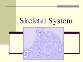
Introduction • How many bones do you think are in the human body? • 206 • Largest Bone? • Femur • Smallest Bone? • Ossicles (ear bones)
1.Support Body Functions (5)
4. Hemopoieses • The bones make blood cells from embryonic month 5 on…
5. Mineral Storage Ca2(PO4)3
Anatomy • There are basically four types of bones.
1. Long Bones e.g. Femur Radius ulna humerus
2. Short bones carpals
Parts of a long bone • Please color code the femur. Color code letters a-g.
A1: Cartilage growth plates on bone ends. (Growth plate) Epiphyseal plate
(hyaline) cartilage on end of bone b bone trabeculae of spongy bone c red marrow cavity d epiphyseal plate (hyaline cartilage)
a Epiphyseal plate made of hyaline cartilage is responsible for long bone growth. Note: The direction of growth is toward the diaphysis (shaft of long bone). Also Note: The newly forming spongy bone (below the growth plate) is not clearly organized as the older spongy bone in the epiphysis above the growth plate.
B. Shaft of the bone, middle part. Diaphysis
C.Cartilage layer to reduce pain and friction. Articular Cartilage
D. Living layer surrounding bone. Nourishing and growth in width. Periosteum
Looks like a sponge. Mostly in the epiphysis Contains red marrow (Makes RBC’s) Spongy bone
Close together in diaphysis. Organized into concentric layers. Compact Bone
Hole in the middle of the bone. Filled with yellow marrow (fat for energy storage) Medullary Cavity
Surface features: (3) • 1. Projections • 2. Depressions • 3. Openings
Projections • For attachments
Depressions • For joints to fit together.
Openings • For blood vessels and nerves.
Osteocyte • Mature bone cells. • Maintain bones and assist and repair.


