Download
1 / 7
90 likes | 249 Views
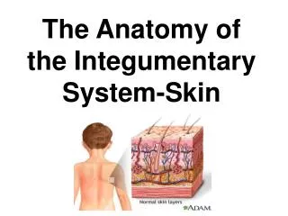
The anatomy of the skin, depth of burns and Jackson burn wound model. JAHD Presentation Perry Crofts. The skin. The largest organ in the body Consists of the epidermis and dermis Epidermis is the outer layer of stratified squamous epithelium. It is avascular and varies in thickness

E N D
The anatomy of the skin, depth of burns and Jackson burn wound model JAHDPresentation Perry Crofts
The skin • The largest organ in the body • Consists of the epidermis and dermis • Epidermis is the outer layer of stratified squamous epithelium. It is avascular and varies in thickness • The dermis is a dense bed of vascular connective tissue • Functions as a physical barrier and a sensory and thermoregulatory organ
Superficial erythema • Only the epidermis involved • Red • Painful • Technically not a burn • Not included in total body surface area calculations
Deep & superficial partial thickness • Superficial partial thickness • Red with clear blisters • Deep partial thickness • Red and white with bloody blisters
Full thickness and Fourth degree burns • Full thickness burns • Extend through entire dermis (full thickness) • Stiff, white and brown • Painless • Fourth Degree burns • Extends through entire skin and into underlying tissue • Black and charred • Painless
Jackson burn wound model • Considers the effect of secondary injury on burns • Zone of coagulation is central, this tissue is most severely damaged and lost • Outside is the zone of stasis which can potentially recover • Most periphery is the zone of hyperaemia