Download
1 / 45
450 likes | 597 Views
Chapter 10: Muscle Tissue. Muscle Tissue. A primary tissue type, divided into : Cardiac muscle Smooth muscle Skeletal muscle Attached to bones Allows us to move Contains CT, nerves and blood vessels. Functions of Skeletal Muscles. 1. 2. 3. 4. 5. CT Organization – 3 layers.

E N D
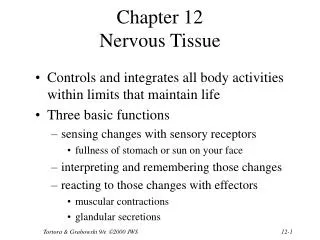
Muscle Tissue • A primary tissue type, divided into: • Cardiac muscle • Smooth muscle • Skeletal muscle • Attached to bones • Allows us to move • Contains CT, nerves and blood vessels
Functions of Skeletal Muscles 1. 2. 3. 4. 5.
CT Organization – 3 layers 1. Epimysium • Surrounds entire muscle • Separates muscle from surrounding tissues • Connected to deep fascia
1. Perimysium • Divides the skeletal muscle into a series of compartments • Each compartment contains a bundle of muscle fibers:
1. Endomysium • Surrounds individual skeletal muscle fibers • Interconnects adjacent muscle fibers • Satellite Cells -
At the end of a muscle: • All 3 layers come together to form a: • Both attach skeletal muscles to bones • Tendon fibers extend into the bone matrix
Skeletal muscle cells are called fibers Enormous Multinucleate Myoblasts fuse during development to form individual skeletal muscle fibers Microanatomy of Skeletal Muscle Fibers
Microanatomy of Skeletal Muscle Fibers • Sarcolemma – cell membrane of muscle fiber • Surround sarcoplasm • Change in the transmembrane potential is the start of a contraction • Transverse Tubules – continuous with sarcolemma and extends into the sarcoplasm • form passageways through muscle fibers • Filled with extracellular fluid • Action potentials
Microanatomy of Skeletal Muscle Fibers • Myofibrils – cylindrical structures encircled by T tubules • As long as the cell • Made of myofilaments • Thin filaments - actin • Thick filaments – myosin • Responsible for muscle fiber contraction • Mitochondria and glycogen
Microanatomy of Skeletal Muscle Fibers • Sarcoplasmic Reticulum – similar to ER of other cells • Forms network around each myofibril • Terminal cisternae – expanded chambers of SR on either side of a T tubule • Ca+2 ions storage • Triad – pair of terminal cisternae plus a T tubule • Separate fluids
Microanatomy of Skeletal Muscle Fibers • Sarcomere – repeating contractile units that make up myofibrils • Smallest functional unit in muscle fibers • Muscle contraction • Made up of: thick and thin filaments, stabilizing proteins and regulating proteins • Striated
Microanatomy of Skeletal Muscle Fibers • A bands – dark bands at center of sarcomere • Thick filaments (myosin) • Contains: • M line – center of A band, connects each thick filament together • H zone – lighter region on either side of M line, contains thick filaments • Zone of overlap – thick and thin filaments overlap one another
Microanatomy of Skeletal Muscle Fibers • I bands – light bands on both sides of A band • Thin filaments (actin) • Contains: • Z lines – boundary between adjacent sarcomeres • Titin – protein that aligns thick and thin filaments • Extends from thick filaments
Level 4: Myofibril Level 1: Skeletal Muscle Level 2: Muscle Fascicle Level 5: Sarcomere Level 3: Muscle Fiber
Muscle Contraction • Sliding Filament Theory • Caused by interactions of thick and thin filaments • Triggered by free Ca2+ in sarcoplasm
Muscle Contraction • Thin Filaments – made of 4 proteins: • F actin – 2 twisted strands of G actin, contain active sites for the binding of myosin • Nebulin – holds 2 strands of G actin together • Tropomyosin – covers G actin active sites to prevent actin/myosin interactions • Troponin – holds tropomyosin to G actin AND contains a site for the binding of Ca2+ • Holds until Ca2+ binds to the active site • Contraction can only occur if position changes
Muscle Contraction • Thick Filaments – consist of a pair of myosin subunits wrapped around each other • Tail – binds to other myosin molecules • Head – 2 subunits, project towards nearest thin filament • During muscle contractions myosin heads pivot towards thin filaments, forming cross-bridges with G actin active sites
Muscle Contraction • Sliding Filament Theory • Thin filaments slide towards M line – shortening • A band remains the same, but the Z lines move closer together
Muscle Contraction • Neuromuscular Junction - NMJ • Where the action potential starts • Each branch ends at a synaptic terminal, which contains mitochondria and Acetylcholine • Neurotransmitter that alters the permeability of the sarcolemma
Muscle Contraction • Synaptic cleft – • Motor end plate – • Both contain AChE – breaks down Ach • Action potential travels along the nerve axon and ends at the synaptic terminal, which changes the permeability • ACh is released
Muscle Contraction • ACh diffuses across the synaptic cleft and binds to ACh receptors on motor end plate • Increase in membrane permeability to sodium ions that rush into the sarcoplasm • Keeps going until AChE removes all ACh • Travels along sarcolemma to T tubules and leads to excitation-contraction coupling - • Action potential leads to contraction • Triads release Ca2+ • Triggers muscle contractions
Muscle Contraction at Sarcomere 1. Exposure of active sites • Calcium ions bind to troponin, changing its position and shifting tropomyosin away from active sites 2. Attachment of cross-bridges • Myosin heads bind to active sites
Muscle Contraction at Sarcomere 3. Pivoting • Power stroke 4. Detachment of cross-bridges • ATP binds to myosin head, link is broken • Attach to another active site
Muscle Contraction at Sarcomere Muscle Contraction at Sarcomere 5. Reactivation of myosin • ATP to ADP and phosphate • Cycle is repeated • All sarcomeres contract at the same time • Contraction duration depends on: • Duration of neural stimulus • Amount of free Ca2+ ions in sarcoplasm • Availability of ATP
Muscle Contraction • 1. At NMJ, ACh is released and binds to receptors on sarcolemma • 2. Change in transmembrane potential results in action potential that spreads across entire surface of cell and T tubules • 3. SR releases stored calcium ions, increasing Ca2+ around sarcomeres • 4. Calcium ions bind to troponin, which exposes active sites on thin filaments and cross-bridges form • 5. Contraction begins as repeated cycles of cross-bridge formation and detachment happen
Muscle Contraction • 6. ACh is broken down by AChE and action potential ends • 7. SR reabsorbs calcium ions and concentration in sarcoplasm decreases • 8. Active sites are re-covered • 9. Contraction ends • 10. Muscle relaxation – sarcomeres remain uncontracted
Rigor Mortis • Stop in blood circulation causes skeletal muscles to be deprived of oxygen and nutrients – • SR becomes unable to pump calcium ions out of sarcoplasm • Extra calcium ions trigger a sustained contraction • Cross-bridges form, but cannot detach • Lasts 15-25 hours after death
2 Types of Muscle Tension • Isotonic Contraction • Skeletal muscle changes length resulting in motion • If muscle tension > resistance: muscle shortens (concentric contraction) • If muscle tension < resistance: muscle lengthens (eccentric contraction)
2 Types of Muscle Contraction • Isometric Contraction • Muscle develops tension, but does not shorten
Resistance and Speed of Contraction • Inversely related • The heavier the resistance on a muscle: • the longer it takes for shortening to begin • the less the muscle will shorten
Muscle Relaxation • After contraction, a muscle fiber returns to resting length by: • Elastic forces • The pull of elastic elements (tendons and ligaments) • Expands the sarcomeres to resting length • Opposing muscle contractions • Reverse the direction of the original motion • The work of opposing skeletal muscle pairs • Gravity • Can take the place of opposing muscle contraction to return a muscle to its resting state
ATP and Muscle Contraction • Muscle contraction uses a lot of ATP • Muscles store enough energy to start contraction, but must manufacture more ATP • Generates ATP at the same rate that it is used • ATP and CP • ATP – active energy model (aerobic and anaerobic) • Creatine Phosphate (CP) – storage molecule for excess ATP in resting muscle • ATP – 2 seconds • CP – 15 seconds • Glycogen – 130 seconds (anaerobic) and 40 mins (aerobic) • Fats
ATP and Muscle Contraction • At rest: • Cells use fatty acids to create CP, ATP and glycogen – rebuilding their storages (beta oxidation) • Moderate Activity: • Cells use fatty acids or glucose and oxygen to produce ATP (aerobic respiration) • Muscle wont fatigue until all energy is used up • Marathon runners • Peak Activity • Cells use oxygen faster than it is supplied • Aerobic resp only provides 1/3 of needed ATP • Anaerobic resp provides the rest – lactic acid
Muscle Fatigue • When muscles can no longer perform a required activity, they are fatigued • Results of Muscle Fatigue: Depletion of metabolic reserves Damage to sarcolemma and SR Low pH (lactic acid) Muscle exhaustion and pain • The Recovery Period • The time required after exertion for muscles to return to normal • Oxygen becomes available • Mitochondrial activity resumes
Muscle Fatigue • The Cori Cycle • The removal and recycling of lactic acid by the liver • Liver converts lactic acid to pyruvic acid • Glucose is released to recharge muscle glycogen reserves Oxygen Debt – after exercise: Body needs more oxygen than usual to normalize metabolic activity Heavy breathing
3 Types of Skeletal Fibers • Fast Fibers: • Contract quickly • High CP • Large diameter, huge glycogen reserves and few mitochondria • Strong contractions, but fatigue quickly • White meat – chicken breast • Slow Fibers • Slow to contract and slow to fatigue • Low CP • Small diameter, but a lot of mitochondria • High oxygen supply • Contain myoglobin (red pigment, binds to oxygen) • Dark meat – chicken legs
3 Types of Muscle Fibers • Intermediate Fibers • Mid-sized • Low myoglobin • More capillaries than fast fibers, slower to fatigue • Table 10-3, page 298 • Human Muscles
Muscle Hypertrophy - muscle Growth from heavy training • increases diameter of muscle fibers • increases number of myofibrils • increases mitochondria, glycogen reserves • Muscle Atrophy – lack of muscle activity • Reduced in muscle size, tone and power
Physical Conditioning • Anaerobic Endurance • Uses fast fibers, fatigues quickly with strenuous activities • 50 m dash, weightlifting • Improved by frequent, brief, intensive workouts – interval training • Aerobic Endurance – supported by mitochondria • Prolonged activity – uses a lot of oxygen and nutrients • Marathon running • Improved by repetitive and cardiovascular training
Cardiac Muscle Tissue • Striated tissue • Smaller cells with single nucleus • Short T-tubules and sarcoplasm • No triads or terminal cisternae • All aerobic • High in myoglobin and mitochondria • Intercalated discs
Smooth Muscle • Blood vessels, reproductive and digestive systems, etc • Different arrangement of actin and myosin • Non-striated