Download

1 / 151
1.64k likes | 3.55k Views
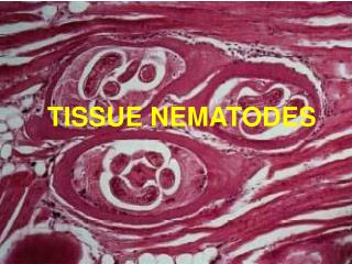
Chapter 10: Muscle Tissue. Muscle Tissue. A primary tissue type, divided into: skeletal muscle Voluntary striated muscle, controlled by nerves of the central nervous system cardiac muscle Involuntary striated muscle smooth muscle Involuntary nonstriated muscle.

E N D
Muscle Tissue • A primary tissue type, divided into: • skeletal muscle • Voluntary striated muscle, controlled by nerves of the central nervous system • cardiac muscle • Involuntary striated muscle • smooth muscle • Involuntary nonstriated muscle
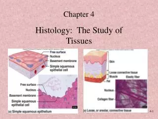
Characteristics of all Muscle Tissues • Specialized Cells: - elongated, high density of myofilaments = cytoplasmic microfilaments of actin and myosin • Excitability/Irritability: - receive and respond to stimulus • Contractility: - shorten and produce force upon stimulation • Extensibility: - can be stretched • Elasticity: - recoil after stretch
Skeletal Muscle Tissue • Skeletal muscles make up 44% of body mass • Skeletal muscle = an organ • composed of: • skeletal muscle cells (fibers) and CT • nerves and blood vessels
Functions of Skeletal Muscles • Produce skeletal movement • Maintain posture and upright position • Support soft tissues • Guard entrances and exits • Maintain body temperature by generating heat • Stabilize joints
Formation of Skeletal Muscle Fibers • Skeletal muscle cells are called fibers Figure 10–2
Skeletal Muscle Anatomy • Each muscle is innervated by one nerve: • Nerve must branch and contact each skeletal muscle fiber (cell) • One artery, branches into extensive capillaries around each fiber: • supply oxygen • supply nutrients • remove wastes.
Organization of Connective Tissues Figure 10–1
Organization of Connective Tissues • Muscles have 3 layers of connective tissues that hold the muscle together: • Epimysium - covers the muscle (exterior collagen layer), separates muscle from other tissues, composed of collagen, connects to deep fascia • Perimysium - composed of collagen and elastin, has associated blood vessels and nerves, bundles muscle fibers into groups called fascicles - perimysium covers a fascicle • Endomysium - composed of reticular fibers, contains capillaries, nerve fibers and satellite cells (= stem cells repair), surrounds individual muscle fibers
Muscle Attachments • Endomysium, perimysium, and epimysium come together: • at ends of muscles • to form connective tissue attachment to bone matrix • Tendon = cord-like bundles • Aponeurosis = sheet-like
How would severing the tendon attached to a muscle affect the muscle’s ability to move a body part? Uncontrolled movement would result from a severed tendon. Movement would be greatly exaggerated with no tendon. No movement is possible without a muscle to bone connection. Limited movement would result.
Skeletal Muscle Fibers • Huge cells: • up to 100 µm diameter, 30 cm long • Multinucleate • Formed by fusion of 100s of myoblasts • Nuclei of each myoblast retained to provide enough mRNA for protein synthesis in large fiber • Unfused myoblasts in adult = satellite cells • Satellite cells are capable of division and fusion to existing fibers for repair but cannot generate new fibers
Organization of Skeletal Muscle Fibers Figure 10–3
Skeletal Muscle Fibers • Cell membrane = sarcolemma • Sarcolemma maintains separation of electrical charges resulting in a transmembrane potential • Na+ pumped out of the cell creating positive charge on the outside of the membrane • Negative charge from proteins on inside give muscle fibers a resting potential of -85mV • If permeability of the membrane is altered, Na+ will flow in causing a change in membrane potential • Change in potential will signal the muscle to contract
Transverse Tubules • Tubes of sarcolemma called transverse tubules (T tubules) reach deep inside the cell to transmit changes in transmembrane potential to structures inside the cell • Transmit action potential through cell • Allow entire muscle fiber to contract simulataneously
Skeletal Muscle Fibers • Cytoplasm = sarcoplasm: • rich in glycosomes (glycogen granules) and myoglobin (binds oxygen) • Fiber is filled with myofibrils extending the whole length of the cell • Myofibrils consist of bundles of myofilaments • Myofilaments are responsible for muscle contraction • made of actin and myosin proteins • 80% of cell volume
Organization of Skeletal Muscle Fibers Figure 10–3
Skeletal Muscle Fibers • Actin: • makes up the thin filament • Myosin: • makes up the thick filament • When thick and thin filaments interact, contraction occurs
Skeletal Muscle Fibers Sarcoplasm contains networks of SER called sarcoplasmic reticulum (SR) • Sarcoplasmic Reticulum: • A membranous structure surrounding each myofibril • Function: • store calcium and help transmit action potential to myofibril • SR forms chambers (terminal cisternae) attached to T-tubules • Cisternae • Concentrate Ca2+ (via ion pumps) • Release Ca2+ into sarcomeres to begin muscle contraction • All calcium is actively pumped from sarcoplasm to SR (SR has 1000X more Ca2+ than sarcoplasm)
Skeletal Muscle Fibers • Triads are located repeated along the length of myofilaments • Triads = T-tubule wrapped around a myofibril sandwiched between two terminal cisternae of SR • Formed by 1 T tubule and 2 terminal cisternae of SR • Triads are located on both ends of a sarcomere • Sarcomere = smallest functional unit of a myofibril
Each muscle = ~ 100 fascicles • Each fascicle = ~ 100 muscle fibers • Each fiber (cell) = ~ 1 thousand myofibrils • Each myofibril = ~ 10 thousand sarcomeres
Sarcomeres • The contractile units of muscle • Structural units of myofibrils • Form visible patterns within myofibrils
Sarcomeres Composed of: 1. Thick filaments – myosin 2. Thin filaments – actin 3. Stabilizing proteins: -hold thick and thin filaments in place 4. Regulatory proteins: - control interactions of thick and thin filaments Organization of the proteins in sarcomere causes striated appearance of the muscle fiber Figure 10–4
Muscle Striations • A striped or striated pattern within myofibrils: • alternating dark, thick filaments(A bands) and light, thin filaments(I bands)
Regions of the Sarcomere • A-band: - whole width of thick filaments, looks dark microscopically • M line: at midline of sarcomere - Center of each thick filament, middle of A-band - Attaches neighboring thick filaments • H-zone: - Light region on either side of the M line - Contains thick filaments only • Zone of overlap: - ends of A-bands - place where thin filaments intercalate between thick filaments (triads encircle zones of overlap)
Regions of the Sarcomere • I-band: - Contains thin filaments outside zone of overlap - Not whole width of thin filaments • Z lines/disc: - the centers of the I bands - constructed of Actinins - Anchor thin filaments and bind neighboring sarcomeres - Constructed of Titin Proteins - Bind thick filaments to Z-line, stabilize the filament
Why does skeletal muscle appear striated when viewed through a microscope? Z lines and myosin filaments align within the tissue. Glycogen reserves are linearly arranged. Capillaries regularly intersect the myofibers. Actin filaments repel stain, appearing banded.
Sarcomere Function • Transverse tubules encircle the sarcomere near zones of overlap • Ca2+ released by SR causes thin and thick filaments to interact • Muscle Contraction • Is caused by interactions of thick and thin filaments • Structures of protein molecules determine interactions
Thin Filament Figure 10–7a
Thin Filaments (5-6 nm diameter) • Made of 4 proteins: • Actin • Nebulin • Holds F actin strands together • F-actin (filamentous) consists of rows of G-actin (globular) • Each G-actin has an active site that can bind to myosin • Tropomyosin - Covers the active sites on G actin to prevent actin–myosin binding • Troponin: holds tropomyosin on the G-actin • Also has receptor for Ca2+: • when Ca2+ binds to the troponin-tropomyosin complex it causes the release of actin allowing it to bind to myosin
Troponin and Tropomyosin Figure 10–7b
Initiating Contraction • Ca2+ binds to receptor on troponin molecule • Troponin–tropomyosin complex changes • Exposes active site of F actin
Thick Filament Figure 10–7c
Thick Filaments (10-12 nm diameter) • Composed of: • bundled myosin molecules • titin strands that recoil after stretching • Each Myosin has three parts 1. Tail: - tails bundled together to make length of thick filament - all point toward M-line 2. Hinge: - flexible region, allows movement for contraction
Thick Filaments (10-12 nm diameter) 3. Head: - hangs off tail by hinge, will bind actin at active site. - No heads in H-zone - also contains core of titin: - elastic protein that attaches thick filaments to Z-line - Titin holds thick filament in place and aid elastic recoil of muscle after stretching - Each thick filament is surrounded by a hexagonal arrangement of thin filaments with which it will interact
The Myosin Molecule Figure 10–7d
Myosin Action • During contraction, myosin heads: • interact with actin filaments, forming cross-bridges • pivot, producing motion
Sliding Filaments Figure 10–8
Sliding Filament Theory Contraction of skeletal muscle is due to thick filaments and thin filament sliding past each other • not compression of the filaments • H-zones and I-bands decrease width during contraction • Zones of overlap increase width • Z-lines move closer together • A-band remains constant Sliding causes shortening of every sarcomere in every myofibril in every fiber Overall result = shortening of whole skeletal muscle
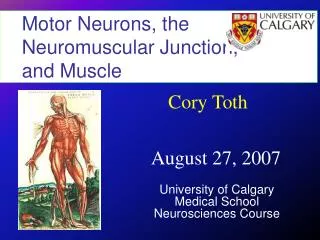
The components of the neuromuscular junction, and the events involved in the neural control of skeletal muscles.
Skeletal Muscle Contraction • Excitation • Excitation-Contraction Coupling • Contraction • Relaxation Figure 10–9 (Navigator)
Excitation and the Neuromuscular Junction • Excitation of muscle fiber is controlled by the nervous system at the neuromuscular junction using neurotransmitter
The Neuromuscular Junction • Is the location of neural stimulation • Action potential (electrical signal): • travels along nerve axon • ends at synaptic terminal
Components of Neuromuscular Junction Neuromuscular Junction: - where a nerve terminal interfaces with a muscle fiber at the motor end plate - one junction per fiber: control of fiber from one neuron • Synaptic Terminal: - expanded end of the axon, contains vesicles of neurotransmitters Acetylcholine (Ach) • Motor End Plate: - specialized sarcolemma that contains Ach receptors and the enzyme acetylcholinesterase (AchE) • Synaptic Cleft: - space between the synaptic terminal and motor end plate where neurotransmitters are released
Skeletal Muscle: Neuromuscular Junction Figure 10–10a, b (Navigator)