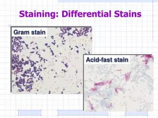
Differential staining
Differential staining. Spore stain Capsule stain. Spore stain.


Differential staining
E N D
Presentation Transcript
Differential staining Spore stain Capsule stain
Spore stain • Members of the anaerobic genera Clostridium species, aerobic Bacillus species are examples of organisms that have the capacity to exist either as metabolically active Vegetative cells or resistant metabolically inactive cells Spores . • Non active, resistant to heating and chemicals. because of the composition of spore layers. • Remain dormant until favorable conditions return.
Not easily stained: • Mild heating. • Use of strong stain. • Timing of the stain is extended.
Spore stain reagents 1-Primary stain: Malachite green, both vegetative cell and spores will appear green. 2-Decolorizing Agent: Water, it will remove the color from the cell, spores will remain green. 3-Counter stain: Safraninwhich will be absorbed by the vegetative cell.
Spore stain steps • Apply malachite green for 5 min application of heat is required. • Rinse with D. water. • Stain with safranin for 1 min. • Observation: • Spores appear green While vegetative bacteria or non spore forming bacteria appear red/pink
Capsule stain • A Capsule is gelatinous outer layer that is secreted by the cell and that surrounds and adheres to the cell wall. Capsules are virulent and capable of producing disease, since the structure protects bacteria against phagocytes of the host cell. • Capsules are non ionic, uncharged because they are composed of polysaccharides.
Capsule stain • Capsule staining is more difficult than other types of differential staining procedures because the capsular material are water-soluble and maybe removed by washing. Smear shouldn’t be heated because the resultant cell shrinkage may create a clear zone around the organism that can be mistaken for the capsule.
This method use Nigrosin and Safranin to stain the bacteria and the background leaving capsule unstained as a clear halo surrounding bacteria.
Capsule stain steps • Deliver 1 drop of Nigrosin by the loop and 1 drop of safranin at the end of clean slide. • Mix a loopful of culture ( Kelbsiella sp.) with the stain. • Spread the mixture on the slide as a blood film. • Air dry the film.