Download

1 / 67
810 likes | 1.55k Views
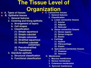
The Tissue Level of Organization . Advanced Anatomy. Epithelial Tissue. Intro to tissues. Overview. No single cell is able to perform the many functions of the human body Instead, through differentiation, each cell specializes to perform a relatively restricted range of functions

E N D
The Tissue Level of Organization Advanced Anatomy
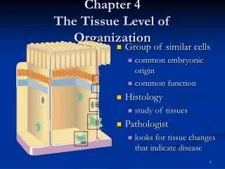
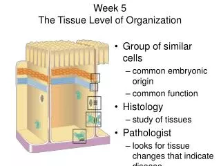
Overview • No single cell is able to perform the many functions of the human body • Instead, through differentiation, each cell specializes to perform a relatively restricted range of functions • Although there are trillions of individual cells in the human body, there are only about 200 different types of cells • These cells combine to form tissues, collections of specialized cells that perform a limited number of functions • Histology is the study of tissues
CLICK IT • The study of tissues is called • A. Microbiology • B. Cellology • C. Histology • D. Tissuology
Epithelial Tissue • Epithelial Tissue includes epithelia and glands • Epithelia are layers of cells that cover internal and external surfaces • Glands are made up of secreting cells derived from epithelia
Characteristics of Epithelia • Cells that are bound closely together • A free surface exposed to the environment or to some internal chamber or passageway • Attachment to underlying connective tissue by a basement membrane • The absence of blood vessels • Because of this avascular condition, epithelial cells must obtain nutrients from deeper tissues or from exposed surfaces
Click It • All of these are characteristics of epithelia except • A. Attachment to the basement membrane • B. A free surface exposed to the environment • C. Vascular • D. Cells that are bound closely together
Functions of the Epithelia • Provide physical protection. Protects exposed and internal surfaces from abrasion, dehydration and destruction by chemical or biological agents • Skin protects against impacts and scrapes, restricts water loss and prevents invasions • Control Permeability. Any substance that enters or leaves the body has to cross the epithelial • Provide sensation. Specialized epithelial cells can detect changed in the environment and relay information about such changes to the nervous system • Touch receptors in the deepest layers of the epithelium of the skin respond by stimulating nerves
Glands • Produce specialized secretions. Epithelial cells that produce secretions are called gland cells. • In a glandular epithelium, most or all of the cells actively produce secretions • These secretions are classified according to their discharge location • Exocrine – Secretions are discharged onto the surface of the skin or other epithelial surface. • Enzymes entering the digestive tract, perspiration on the skin, milk produced by mammary glands • Endocrine – Secretions are released into the surrounding tissues and blood • Hormones
Concept Check • What are the four essential functions of epithelial tissue? • Provide physical protection • Control Permeability • Provide sensation • Produce specialized secretions
Intercellular Connections • To be effective in protecting other tissues, epithelial cells must remain firmly attached to one another • When the epithelium of the skin is damaged by a burn or abrasion, disease-causing bacteria can enter the underlying tissues and cause infection • Undamaged epithelia form effective barriers because the epithelial cell membranes are held together by an intercellular cement and by specialized attachment sites called cell junctions • Types: Tight junction, gap junctions and desmosomes
Tight Junction • At a tight junction the outermost lipid layers of adjacent cell membranes are tightly bound together by interlocking membrane proteins • Tight junctions prevent the passage of water and solutes between the cells. • The junctions are common between epithelial cells exposed to harsh chemicals or powerful enzymes • Example: tight junctions between cells lining the digestive tract keep digestive enzymes, stomach acids or waste from damaging underlying tissues
Gap Junctions • At a gap junction, two cells are held together by interlocked membrane proteins • Because these are channel proteins, the result is a narrow passageway that lets small molecules and ions pass from cell to cell • Gap junctions interconnect cells in some epithelia, but they are most abundant in cardiac muscle and smooth muscle tissue
Desmosomes • At desmosomes, the cell membranes of two cells are locked together by intercellular cement and a network of protein filaments. • A desmosome may form small discs or encircle a cell • Desmosomes are very strong, and the connection can resist stretching and twisting. • In the skin, these links are so strong that dead cells are usually shed in sheets, rather than individually
Click It • The most abundant connections between cells in the superficial layers of the skin are • A. Intermediate Junctions • B. Gab Junctions • C. Desmosomes • D. Tight Junctions
The Epithelial Surface • Many epithelia that line internal passageways have microvilli on their exposed surfaces. • They may vary from just a few to so many that they carpet the surface • Microvilli are especially abundant on epithelial surfaces where absorption and secretion takes place • Digestive and urinary tracts • The epithelial specialize in the active and passive transport of materials across the cell membrane • Some epithelia contain cilia on their exposed surfaces • Respiratory tract
Click It • Which of the following tissues is avascular • A. Cardiac • B. Stratified squamous epithelial • C. Compact Bone • D. Skeletal
The Basement Membrane • Epithelial cells not only must hold onto one another, byt also must remain firmly connected to the rest of the body • This function is performed by the basement membrane, which lies between the epithelium and connective tissues • There are no cells within the basement membrane, which consists of a network of protein fibers. • Epithelial cells adjacent to the basement membrane are firmly attached to the proteins by hemidesmosomes
Epithelial renewal and repair • An epithelium must continually repair and renew itself • Epithelial cells may survive for just a day or two, because they are lost or destroyed by exposure to destructive enzymes, toxic chemicals, pathogenic bacteria, or mechanical abrasion. • The only way epithelium can maintain its structure over time is through the continual division of stem cells • These cells are found in the deepest layers of the epithelium, near the basement membrane
Classifying Epithelia • Epithelia are classified according to the number of cell layers and the shape of the exposed cells • This classification scheme recognizes two types of layering – simple and stratified – and three cell shapes – squamous, cubodial and columnar
Click It • Epithelial tissue is classified by • A. Number of Layers • B. Composition of Matrix • C. Cell Shape • D. Both the number of layers and the cell shape
Cell Layers • A simple epithelium consists of a single layer of cells covering the basement membrane • Simple epithelia are thin • Since a single layer of cells is fragile and cannot provide much mechanical protection, it is only found in the protected areas inside the body • They line internal compartments and passageways, including body cavities and the interior of the heart and blood vessels • Characteristic of regions where secretion and absorption occur and the gas exchange surfaces of the lungs
Stratified Epithelium • Stratified epithelium provides a greater degree of protection because it has several layers of cells above the basement membrane • Stratified epithelia are usually found in areas subject to mechanical or chemical stresses, such as the surface of the skin and the linings of the mouth and anus.
Click It • A tissue that is one layer thick but appears to be multilayered and is composed of cells taller than they are wide is. (Hint: What does Psuedo mean) • A. Stratified ciliated columnar • B. Simple Squamous • C. Pseduostratified Columnar • D. Transitional
Cell shape • In sectional view, the cells at the surface of the epithelium have one of three basic shapes • Squamous – the cells are thin and flat and the nucleus occupies the thickest portion of each cell. Viewed from the surface it looks like fried eggs • Cuboidal – resemble little hexagonal boxes when seen in three dimensions, but in typical sectional view they appear square. The nuclei lie near the center of each cell and they form a neat row • Columnar – the cells are also hexagonal, but taller and more slender. The nuclei are crowded into a narrow band close to the basement membrane
Simple Squamous Epithelia • A simple squamous epithelium is found in protected regions here absorption takes place or where a slick, slippery surface reduces friction • Examples: portion of the kidney tubules, the exchange surfaces of the lungs, the lining of body cavities, and the lining of blood vessels and the heart
Simple Cuboidal Epithelia • Simple Cuboidal Epithelia provides limited protection and occurs in regions where secretion or absorption take place • These functions are enhanced by larger cells that have more room for the necessary organelles. • Simple cuboidal epithelia secrete enzymes and buffers in the pancreas and salivary glands and line the ducts that discharge these secretions • Simple cuboidal epithelia also line portions of the kidneys involved in urine production
Simple Columnar Epithelia • Simple Columnar Epithelia provides some protection and may also occur in areas of absorption and secretion. • This type of epithelium lines the stomach, the intestinal tract and many excretory ducts
Transitional Epithelia • Transitional Epithelia withstands considerable stretching. • It lines the ureters and urinary bladder, where large changes in volume occur • In an empty bladder, the epithelium seems to have many layers, and the outermost cells appear rounded. • The layered appearance results from overcrowding: the actual structure of the epithelium can be seen in the full urinary bladder, when the volume of urine has stretched the lining to its natural thickness
Stratified Squamous Epithelia • Stratified Squamous Epithelia is found where mechanical stresses are severe. • The surface of the skin and the lining of the mouth, tongue, esophagus and anus are good examples.
Glandular Epithelia • Many epithelia contain gland cells that produce exocrine or endocrine secretions • Exocrine secretions are produced by exocrine glands that discharge their products through a duct • Endocrine secretions (hormones) are produced by ductless glands and released into the blood or tissue fluid
Mode of Secretion • A glandular epithelial cell may use one of three methods to release its secretions • Merocrine secretion – the product is released from secretory vesicles by exocytosis • Apocrine secretion – involves the loss of both cytoplasm and the secretory product. The outermost portion of the cytoplasm becomes packed with secretory vesicles before it is shed • Milk production involves both merocrine and apocrine • Holocrine secretion – the entire cell becomes packed with secretions and then bursts apart and dies
Concept Checks • You look at a tissue under a microscope and see a simple squamous epithelium. Can it be a sample of the skin surface? • No. A simple squamous epithelium does not provide enough protection against infection, abrasion, and dehydration and is not found in the skin surface
Concept Check • Secretory cells associated with hair follicles fill with secretions and then rupture, releasing their contents. What type of secretion is this? • Holocrine secretion
Concept Check • What physiological functions are enhanced by epithelial cells bearing microvilli and cilia? • The presence of microvilli on the free surface of epithelial cells greatly increase the surface area for absorption. Cilia function to move materials over the surface of epithelial cells.
Connective Tissue • Connective tissues are the most diverse of the body • All connective tissues have three basic components • Specialized Cells • Protein Fibers • Ground Substance • The extracellular protein fibers and ground substance constitute the matrix that surrounds the cells • Connective tissues are distributed throughout the body but are never exposed to the outside environment • Most connective tissues are highly vascular
Click It • All of these are components of connective tissue except • A. Avascular • B. Specialized Cells • C. Protein Fibers • D. Ground Substance
Functions • Supporting and Protecting • The minerals and fibers produced by connective tissue cells establish a bony structural framework for the body and support organs • Transporting Materials • Fluid connective tissue provides an efficient means of moving dissolved materials • Storing energy reserves • Fats are stored in connective tissue cells called adipose cells until needed • Defending the body • Specialized connective tissue cells respond to invasions by microorganisms through the production of antibodies
Click It • Fats are stored in • A. Blood Cells • B. Lymphocytes • C. Keratin • D. Adipose
Classifying Connective tissue • Connective tissue proper • Loose • Fibers create loose, open framework • Dense • Fibers densely packed • Fluid Connective Tissue • Blood • Lymph • Supporting Connective Tissue • Cartilage • Bone
Click it • The two fluid connective tissues are • A. Cilia and flagella • B. Cartilage and Bone • C. Loose and Dense • D. Blood and Lymph
Connective Tissue Proper Cell Types • Contains varied cell populations, fibers and a syrupy ground substance • Fibroblasts – the most abundant cells in the CTP. • Responsible for the production and maintenance of connective tissue fibers and ground substance • Macrophages – are scattered among fibers • Phagocytize damaged cells or pathogens • Macrophages that spend long periods of time in tissue are called fixed. When an infection occurs, additional macrophages are drawn to infected area
Connective Tissue Proper Cell Types • Fat Cells are known as adipose cells or adipocytes. • Contains such a large droplet of lipid that the nucleus is squeezed to one side of the cell • Varies from one tissue to another, from one region of the body to another and from individual to individual • Mast Cells • Small mobile cells found near blood vessels • Cytoplasm packed with vesicles filled with chemicals that are released to begin the body’s defensive activities after an injury infection
Connective Tissue Fibers • Collagen fibers are long, straight, and unbranched. • Most common in connective tissue • Strong but flexible • Elastic Fibers • Contain Elastic • Branched and wavy • After stretching they will return to original length • Reticular Fibers • Least common of the three • Form branching interwoven framework in various organs
Ground Substance • Ground substance fills the spaces between cells and surrounds the connective tissue fibers • It is clear, colorless and similar in consistency to maple syrup • The consistency slows the movement of bacteria and other pathogens, making them easier for the phagocytes to catch
Categorization • Loose connective tissue and dense connective tissues • Loose connective tissues are the packing material for the body • These tissues fill spaces between organs, provide cushioning, and support epithelia • Dense connective tissues are tough, strong and durable • They resist tension and distortion and interconnect bones and muscles • Form thick fibrous layer called a capsule, that surrounds internal organs, and encloses joint cavities
Loose Connective Tissue • Least specialized connective tissue in the adult body • It contains all of the cells and fibers found in any connective tissue, in addition to an extensive circulatory supply • Forms a layer that separates skin from underlying muscles • Provides the metabolic needs of nearby epithelia tissue
Adipose Tissue • Loose connective tissue containing large numbers of fat cells • The difference between loose connective tissue and adipose tissue is one of degree – loose connective tissue is called adipose when it becomes dominated by fat cells • It is common under the skin of the sides, buttocks, and breasts. It fills the bony sockets behind the eyes, surrounds the kidneys, and dominates extensive areas of loose connective tissues in the pericardial and peritoneal (abdominal) cavities