Download
1 / 33
380 likes | 1.47k Views
A. Types of tissues B. Epithelial tissues 1. General features 2. Covering and lining epithelia a. Arrangement of layers b. Cell shapes c. Classification (1) Simple squamous (2) Simple cuboidal

E N D
A. Types of tissues B. Epithelial tissues 1. General features 2. Covering and lining epithelia a. Arrangement of layers b. Cell shapes c. Classification (1) Simple squamous (2) Simple cuboidal (3) Simple columnar (4) Stratified squamous (5) Stratified cuboidal (columnar) (6) Pseudostratified (7) Transitional 3. Glandular epithelia a. Structural classification b. Functional classification C. Connective tissues 1. General features 2. Classification a. Loose connective tissues (1) Areolar (2) Adipose (3) Reticular b. Dense connective tissues (1) Dense regular (2) Dense irregular (3) Elastic c. Cartilage (1) Hyaline (2) Fibrocartilage (3) Elastic d. Specialized connective tissues (1) Osseous (2) Vascular D. Membranes 1. Mucous membranes 2. Serous membranes 3. Cutaneous membranes 4. Synovial membranes The Tissue Level of Organization
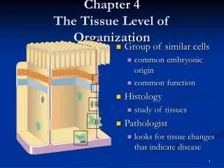
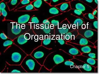
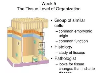
What is a tissue? • A similar group of cells working together to serve a particular function
Types of Tissue • 1. epithelial • 2. connective • 3. muscle • 4. nervous
Epithelial Tissues • 1. covering and lining • 2. glandular
General Features of Epithelia • 1. closely packed cells • 2. continuous sheets • 3. apical vs basal surface • 4. basement membrane • 5. avascular • 6. nerve supply • 7. high mitotic rate Apical Surface Basal Lamina Vs. Reticular Lamina
Covering and lining epithelia You are responsible for Handout #2 Epithelial Tissues • 1. arrangement of layers a. simple b. stratified c. pseudostratified • 2. shape of apical cells a. squamous b. cuboidal c. columnar d. transitional
Simple Squamous External Surface (Serosa) of Small Intestine Location: lining heart (endocardium) and blood vessels (endothelium), lymphatic vessels, alveoli of lungs, glomerular capsule of kidneys, part of serous membranes Function: diffusion, osmosis, and filtration
Stratified Squamous Skin: Sole of Foot Mucosa of Vagina nonkeratinized forms line mouth and tongue, pharynx, esophagus, anal canal, and vagina Location: keratinized form forms epidermis Function: protection against wear-and-tear
Simple Cuboidal Kidney Tubules Location: lines kidney tubules and small ducts of many glands, covers ovary, forms pigmented epithelium of retina Function: absorption and secretion
Stratified Cuboidal Duct of a Sweat Gland Location: relatively rare; lines larger ducts of some glands and part of the male urethra Function: protection
Simple Columnar Internal Surface (Mucosa) of Small Intestine Location: lines GI tract from stomach to anal canal, ducts of some glands, gallbladder; ciliated form lines oviducts, uterus, central canal of spinal cord Function: Secretion and absorption
Stratified Columnar Location: Limited regions of the Pharynx, larynx, Anal canal, and Male urethra (very rare)
Pseudostratified Mucosa of the Trachea Location: lines much of lower respiratory system down to bronchiolar level; nonciliated form may be found in some gland ducts, epididymis, and part of male urethra Function: secretion from goblet cells, movement of mucous across surface by ciliary action
Transitional Allantoic Duct of Umbilical Cord Location: urinary bladder!!!, portions of ureters and urethra Function: allows distention of organ without causing an increase in tension in wall of organ
Glandular Epithelium • 1. secretory portions of glands • 2. endocrine vs exocrine
Structural classification of exocrine glands • 1. unicellular gland (the goblet cell) • 2. multicellular gland
Functional Classification of Exocrine Glands • 1. merocrine (eccrine) • 2. holocrine • 3. apocrine
Connective Tissues • Three basic elements: 1. cells 2. ground substance 3. fibers a. collagen b. elastin c. reticular fibers • Matrix = ground substance + fiber(s)
Classification of Connective Tissues • 1. Loose connective tissues a. areolar (fibroblastic cell type) b. adipose (adipocytic cell type) c. reticular (fibroblastic cell type) 2. Dense connective tissues a. dense regular (fibroblastic cell type) b. dense irregular (fibroblastic cell type) c. elastic (fibroblastic cell type)
Classification of Connective Tissues Continued… • 3. Cartilage (chondrocytic cell type) a. hyaline b. fibrocartilage c. elastic • 4. Bone • 5. Blood
Know Handout #3 Connective Tissue TypesThree Basic Elements • 1. Cells • Ground substance (ground substance is a more or less homogeneous, amorphous, water-based background substance in which the specific differentiated elements of a connective tissue are suspended. • Fibers (E.g. collagen, elastin, reticular) Matrix= Ground substance + fibers that separates cells
Areolar (Loose) Location(s): papillary dermis of skin, hypodermis, mucous membranes, blood vessels, nerves, and around body organs (i.e. everywhere) Description: loosely woven fibers embedded in ground substance; many different cell types wander through; fibroblasts form basic tissue Function: strength, elasticity, and support
Adipose (Loose) Location: hypodermis; around heart, kidneys, and eyes; yellow bone marrow; around joints Description: adipocytes that store fats, forming a characteristic “signet ring” appearance Function: provide insulation, energy storage, protection from mechanical injury
Reticular (Loose) Location: stroma of liver, spleen, lymph nodes; red bone marrow; basement membranes Description: net work of very short interlacing collagen fibers and fibroblasts Function: form framework of organs, binds together smooth muscle cells
Dense Regular Location: tendons, aponeuroses, and most ligaments Description: collagen fibers arranged in parallel bundles, with fibroblasts scattered between Function: provides strong attachments between parts
Dense Irregular Location: dense fascia, reticular dermis, perichondrium, periosteum, joint capsules, dura mater, membrane capsules, heart valves Description: collagen fibers randomly arranged and fibroblasts, forming a sheet Function: strength, support, and protection
Elastic (Dense) Location: lung, elastic arteries, trachea and bronchial tree, true vocal cords, vertebral ligaments, suspensory ligament of penis Description: elastic fibers that branch freely, forming sheets or ligaments Function: provides extensibility and elasticity to various organs that must be stretched
Hyaline Cartilage Location: articular surfaces of bones; anterior ribs; nose, parts or larynx, trachea, and bronchial tree; embryonic skeleton Description: Chondrocytes embedded in cartilage matrix rich in collagen fibers Function: resists compressive forces, provides smooth surfaces for articulation at synovial joints
Fibrocartilage Location: pubic symphysis, intervertebral discs, menisci of shoulders and knees Description: consists of scattered Chondrocytes with hyaline cartilage matrix among bundles of collagen fibers Function: support, fusion (between pubic bones and between vertebrae), deepening of shoulder and knee joints
Elastic Cartilage Location: epiglottis, external ear, auditory tubes Description: Chondrocytes embedded in cartilage matrix rich in network of elastic fibers Function: allows structure to be semi-rigid and extensible; structure can return to resting shape
Bone Location: comprises the bones of the skeleton Description: osteocytes embedded in a mineralized matrix of collagen fibers and ground substance Function: support, protection, storage, provides levers for movement; provides for blood cell formation
Blood Location: within blood vessels and heart Description: formed cellular elements suspended in a liquid matrix (plasma) Function: transport of gases, immune function, blood clotting
Membranes • 1. mucous membrane (mucosa) • 2. serous membrane (serosa) a. parietal layer vs. visceral layer (serous fluid) b. endothelium (endocardium) c. mesothelium • 3. cutaneous membrane (epidermis) ________________________________________ • 4. synovial membrane (not really a membrane)