Download
1 / 79
790 likes | 865 Views
Explore the four major tissue types' functions and structure, from epithelial to neural tissue, highlighting their roles in the body's organization and function.

E N D
Learning Objectives • Identify the four major tissue types and describe their functions. • Describe the relationship between form and function for each tissue type. • Discuss the types and functions of epithelial tissues. • Compare the structure and function of connective tissues.
Learning Objectives • Explain the structure and function of the four types of membrane. • Describe the three types of muscle tissue and the structural features of each. • Discuss the basic structure and role of neural tissue.
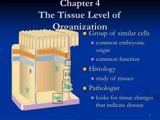
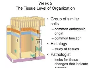
Tissues and tissue types • Tissues are: • Collections of specialized cells and cell products organized to perform a limited number of functions • Histology = study of tissues • The four tissue types are: • Epithelial • Connective • Muscular • Nervous
Epithelial tissue • Includes glands and epithelium • Glands are secretory • Is avascular • Forms a protective barrier that regulates permeability • Cells may show polarity
Functions of epithelium • Physical protection • Control permeability • Provide sensation • Produce specialized secretions
Specializations of epithelium • Perform secretory functions • Perform transport functions • Maintain physical integrity • Ciliated epithelia move materials across their surface
Figure 4.1 The Polarity of Epithelial Cells Figure 4.1
Maintaining the integrity of epithelium • Cells attach via cell adhesion molecules (CAM) • Cells attach at specialized cell junctions • Tight junctions • Desmosomes • Gap junctions
Figure 4.2 Intercellular connections PLAY Animation: Intercellular connections Figure 4.2
Structure of typical epithelium • Basal lamina attaches to underlying surface • Lamina lucida • Lamina densa • Germinative cells replace short-lived epithelial cells
Classification of epithelia • Number of cell layers • Simple • Stratified • Shape of apical surface cells • Squamous • Cuboidal • Columnar
Figure 4.3 Squamous Epithelia Figure 4.3
Figure 4.4 Cuboidal Epithelia Figure 4.4a
Figure 4.4 Cuboidal Epithelia Figure 4.4b
Figure 4.4 Transitional Epithelium Figure 4.4c
Figure 4.5 Columnar Epithelia Figure 4.5a
Figure 4.5 Columnar Epithelia Figure 4.5b
Figure 4.5 Columnar Epithelia Figure 4.5c
Glandular epithelia • Exocrine glands • Secrete through ducts onto the surface of the gland • Endocrine glands • Release hormones into surrounding fluid
Glandular secretions can be: • Merocrine (product released through exocytosis) • Apocrine (involves the loss of both product and cytoplasm) • Holocrine (destroys the cell)
Figure 4.6 Mechanisms of Glandular Secretion Animation: Mechanisms of glandular secretion PLAY Figure 4.6
Glands • Unicellular • Individual secretory cells • Multicellular • Organs containing glandular epithelium • Classified according to structure
Figure 4.7 A Structural Classification of Exocrine Glands Figure 4.7
Connective tissue functions: • Establishing a structural framework • Transporting fluids and dissolved materials • Protecting delicate organs • Supporting, surrounding and interconnecting tissues • Storing energy reserves • Defending the body from microorganisms
Figure 4.8 A Classification of Connective Tissues Figure 4.8
Connective tissues contain • Specialized cells • Matrix • Composed of extracellular protein fibers and a ground substance
Connective tissue proper • Contains varied cell populations • Contains various fiber types • A syrupy ground substance
Fluid connective tissue • Contains a distinctive cell population • Watery ground substance with dissolved proteins • Two types • Blood • Lymph
Supporting connective tissues • Less diverse cell population • Dense ground substance • Closely packed fibers • Two types • Cartilage • Bone
Connective tissue proper • Contains fibers, a viscous ground substance, and a varied cell population • Fibroblasts • Macrophage • Adipocytes • Mesenchymal cells • Melanocytes • Mast cells • Lymphocytes • Microphages
Connective tissue proper • Three types of fiber • Collagen fibers • Reticular fibers • Elastic fibers
Connective tissue proper • Classified as loose or dense • Loose • Embryonic mesenchyme, mucous connective tissues • Areolar tissue • Adipose tissue • Reticular tissue • Dense • Dense regular CT • Dense irregular CT
Figure 4.9 The Cells and Fibers of Connective Tissue Proper Figure 4.9
Figure 4.10 Connective Tissue in Embryos Figure 4.10
Figure 4.11 Adipose and Reticular Tissues Figure 4.11
Figure 4.12 Dense Connective Tissues Figure 4.12a
Figure 4.12 Dense Connective Tissues Figure 4.12b
Figure 4.12 Dense Connective Tissues Figure 4.12c
Fluid connective tissues • Distinctive collections of cells in a fluid matrix • Blood • Formed elements and plasma • Red blood cells, white blood cells and platelets • Arteries carry blood away, veins carry to the heart • Capillaries allow diffusion into the interstitial fluid • Lymph • Interstitial fluid entering the lymphatic vessels
Figure 4.13 Formed Elements of the Blood Figure 4.13
Supporting connective tissues • Cartilage and bone support the rest of the body • Cartilage • Grows via interstitial and appositional growth • Matrix is a firm gel containing chondroitin sulfate • Cells called chondrocytes • Cells found in lacunae • Perichondrium separates cartilage from surrounding tissues • Three types: hyaline, elastic and fibrocartilage
Figure 4.15 The Perichondrium and Types of Cartilage Figure 4.15a, b
Figure 4.15 The Perichondrium and Types of Cartilage Figure 4.15c
Figure 4.15 The Perichondrium and Types of Cartilage Figure 4.15d
Bone, or osseus tissue • Has osteocytes • Depend on diffusion through canaliculi for nutrients • Little ground substance • Dense mineralized matrix • Surrounded by periosteum
Figure 4.16 Bone Figure 4.16