Download
1 / 10
120 likes | 1.05k Views
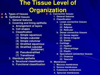
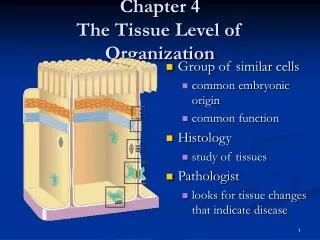
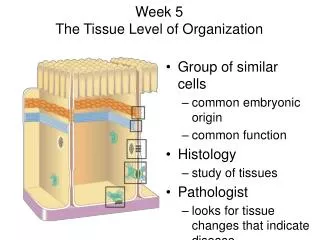
Tissue Level of Organization. Lesson 1. Types of Tissues. Epithelial tissue Covers body surfaces and lines hollow organs, body cavities, and ducts. It also forms glands. Connective tissue Protects and supports the body and its organs.

E N D
Tissue Level of Organization Lesson 1
Types of Tissues • Epithelial tissue • Covers body surfaces and lines hollow organs, body cavities, and ducts. It also forms glands. • Connective tissue • Protects and supports the body and its organs. • Various types of connective tissue bind organs together, store energy reserves, and help provide immunity.
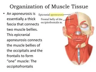
Muscular tissue • Generates the physical force needed for movement • Consists of fibers that are modified for contraction and provide motion, maintenance of posture, and heat • Types of muscle include • Skeletal muscle: attached to bones, is striated, and is voluntary • Cardiac muscle: forms most of the heart wall, is striated, and is usually involuntary • Visceral (smooth) muscle: found in the walls of hollow internal structures, is nonstriated, and is usually involuntary
Nervous tissue • Detects changes in a variety of conditions inside and outside the body and responds by generating action potentials that help maintain homeostasis • Composed of neurons (nerve cells) and neuroglia • Most neurons are composed of a cell body, dendrites, and axons • Neurons are sensitive to stimuli, convert stimuli into nerve impulses, and conduct nerve impulses to other neurons, muscle fibers, or glands • Neuroglia protect and support neurons and are often the sites of tumors of the nervous system
Primary Germ Layers • Tissues of the body develop from three primary germ layers formed in the embryo: ectoderm, mesoderm, and endoderm. • Epithelial tissues develop from all three germ layers. • Connective tissues and most muscle tissues derive from mesoderm. • Nervous tissue develops from ectoderm.
Cell Junctions • Cell junctions are contact points between the plasma membranes of tissue cells. • Tight junctions: consists of weblike strands of transmembrane proteins that fuse the outer surfaces of adjacent plasma membranes together. • Cells of the epithelial tissues that line the stomach, intestines, and urinary bladder have many tight junctions.
Adherens junctions: contain plaque, a dense layer of proteins on the inside of the plasma membrane that attaches to both membrane proteins and to microfilaments of the cytoskeleton. • Transmembrane glycoproteins called cadherins join the cells by inserting into the plaque from the opposite side of the plasma membrane, partially crosses the intercellular space and connects to cadherins of an adjacent cell. • Adherens junctions help epithelial surfaces resist separation during various contractile activities, such as when food moves through the intestines.
Desmosomes: also contain plaque and cadherins. However, the plaque attaches to intermediate filaments, not microfilaments, that contain keratin. The intermediate filaments stretch across the cytoplasma and provide internal support for the cell. • Desmosomes are common in the epidermis cells and cardiac muscle cells where they keep the cells from separating under tension and contraction.
Hemidesmosomes: consist of transmembrane glycoproteins called integrins that attach to intermediate filaments on the inside of the plasma membrane while attaching to laminin on the outside of the plasma membrane. • Hemidesmosomes anchor cells to the basement membrane. • Basement membrane is a layer of collagen, glycoproteins, proteglycans, and fibrous proteins between the epithelium cells and the connective tissue cells.
Gap junctions: the plasma membranes of adjoining cells are not fused together but are separated by a very narrow intercellular gap. • Membrane proteins called connexins form tiny fluid-filled tunnels called connexons that connect neighboring cells. • Through connexons, ions and small molecules can diffuse from the cytoplasm of one cell to another. • Gap junctions also allow cells in a tissue to communicate with each other, enable nerve or muscle impulses to spread rapidly among cells, and allow chemical and electrical signals that regulate growth to pass from cell to cell.