Download
1 / 89
900 likes | 1.43k Views
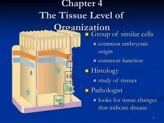
4 The Tissue Level of Organization. An Introduction to Tissues. Tissues Structures with discrete structural and functional properties Tissues in combination form organs, such as the heart or liver Organs can be grouped into 11 organ systems. 4-1 Four Types of Tissue. Tissue

E N D
4 The Tissue Level of Organization
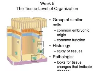
An Introduction to Tissues Tissues Structures with discrete structural and functional properties Tissues in combination form organs, such as the heart or liver Organs can be grouped into 11 organ systems
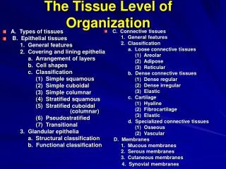
4-1 Four Types of Tissue • Tissue • Are collections of cells and cell products that perform specific, limited functions • Four types of tissue • Epithelial tissue • Connective tissue • Muscle tissue • Neural tissue
4-1 Four Types of Tissue • Epithelial Tissue • Covers exposed surfaces • Lines internal passageways • Forms glands
Connective Tissue • Fills internal spaces • Supports other tissues • Transports materials • Stores energy
4-1 Four Types of Tissue • Muscle Tissue • Specialized for contraction • Skeletal muscle, heart muscle, and walls of hollow organs
Neural Tissue • Carries electrical signals from one part of the body to another
4-2 Epithelial Tissue • Epithelia • Layers of cells covering internal or external surfaces • Glands • Structures that produce secretions
Figure 4-1 The Polarity of Epithelial Cells Cilia Microvilli Apicalsurface Golgiapparatus Nucleus Mitochondria Basement membrane Basolateralsurfaces
4-2 Epithelial Tissue • Functions of Epithelial Tissue • Provide Physical Protection • Control Permeability • Provide Sensation • Produce Specialized Secretions (glandular epithelium)
4-2 Epithelial Tissue • Intercellular Connections • Cell junctions • Form bonds with other cells or extracellular material • Tight junctions • Gap junctions
4-2 Epithelial Tissue • Tight Junctions • Between two plasma membranes • Prevents passage of water and solutes • Isolates wastes in the lumen
Gap Junctions • Allow rapid communication • Are held together by channel proteins (junctional proteins, connexons) • Allow ions to pass • Coordinate contractions in heart muscle
4-3 Classification of Epithelia • Singular = Epithelium; Plural = Epithelia • Classes of Epithelia • Based on shape • Squamous epithelia — thin and flat • Cuboidal epithelia — square shaped • Columnar epithelia — tall, slender rectangles • Based on layers • Simple epithelium — single layer of cells • Stratified epithelium — several layers of cells
4-3 Classification of Epithelia • Squamous Epithelia • Simple squamous epithelium • Absorption and diffusion • Mesothelium • Lines body cavities • Endothelium • Lines heart and blood vessels
Figure 4-3a Squamous Epithelia Simple Squamous Epithelium LOCATIONS: Mesothelia lining ventral body cavities; endothelia lining heartand blood vessels; portions of kidney tubules (thin sections of nephron loops); inner lining of cornea; alveoli of lungs FUNCTIONS: Reduces friction; controls vessel permeability; performsabsorption and secretion Cytoplasm Nucleus Connective tissue LM 238 Lining of peritoneal cavity
4-3 Classification of Epithelia • Squamous Epithelia • Stratified squamous epithelium • Protects against attacks • Keratin protein adds strength and water resistance
Figure 4-3b Squamous Epithelia Stratified Squamous Epithelium LOCATIONS: Surface of skin; lining of mouth, throat, esophagus, rectum, anus, and vagina FUNCTIONS: Provides physical protection against abrasion, pathogens, and chemical attack Squamoussuperficial cells Stem cells Basementmembrane Connectivetissue Surface of tongue LM 310
4-3 Classification of Epithelia • Cuboidal Epithelia • Simple cuboidal epithelium • Secretion and absorption • Stratified cuboidal epithelia • Sweat ducts and mammary ducts
Figure 4-4a Cuboidal and Transitional Epithelia Simple Cuboidal Epithelium LOCATIONS: Glands; ducts;portions of kidney tubules; thyroidgland Connectivetissue FUNCTIONS: Limited protection,secretion, absorption Nucleus Cuboidalcells Basementmembrane Kidney tubule LM 650
Figure 4-4b Cuboidal and Transitional Epithelia Stratified Cuboidal Epithelium LOCATIONS: Lining of some ducts(rare) FUNCTIONS: Protection, secretion,absorption Lumenof duct Stratifiedcuboidalcells Basementmembrane Nuclei Connectivetissue Sweat gland duct LM 500
4-3 Classification of Epithelia • Transitional Epithelium • Tolerates repeated cycles of stretching and recoiling and returns to its previous shape without damage • Appearance changes as stretching occurs • Situated in regions of the urinary system (e.g., urinary bladder)
Figure 4-4c Cuboidal and Transitional Epithelia Transitional Epithelium LOCATIONS: Urinarybladder; renal pelvis;ureters FUNCTIONS: Permitsexpansion and recoilafter stretching Epithelium(relaxed) Basement membrane Connective tissue andsmooth muscle layers LM 400 Empty bladder Epithelium(stretched) Basement membrane LM 400 Connective tissue andsmooth muscle layers LM 400 Full bladder Urinary bladder
4-3 Classification of Epithelia • Columnar Epithelia • Simple columnar epithelium • Absorption and secretion • Pseudostratified columnar epithelium • Cilia movement • Stratified columnar epithelium • Protection
Figure 4-5a Columnar Epithelia Simple Columnar Epithelium LOCATIONS: Lining ofstomach, intestine, gallbladder,uterine tubes, and collectingducts of kidneys Microvilli Cytoplasm FUNCTIONS: Protection,secretion, absorption Nucleus Basementmembrane Looseconnective tissue LM 350 Intestinal lining
Figure 4-5b Columnar Epithelia Pseudostratified Ciliated Columnar Epithelium LOCATIONS: Lining ofnasal cavity, trachea, andbronchi; portions of malereproductive tract Cilia Cytoplasm FUNCTIONS: Protection,secretion, move mucuswith cilia Nuclei Basementmembrane Looseconnective tissue Trachea LM 350
Figure 4-5c Columnar Epithelia Stratified Columnar Epithelium LOCATIONS: Small areas ofthe pharynx, epiglottis, anus,mammary glands, salivarygland ducts, and urethra Looseconnective tissue Deeper basalcells FUNCTION: Protection Superficialcolumnar cells Lumen Lumen Cytoplasm Nuclei Basementmembrane Salivary gland duct LM 175
4-3 Classification of Epithelia • Glandular Epithelia • Endocrine glands • Release hormones • Into interstitial fluid • No ducts • Exocrine glands • Produce secretions • Onto epithelial surfaces • Through ducts
4-3 Classification of Epithelia • Glandular Epithelia • Types of Secretions • Serous glands • Watery secretions • Mucous glands • Secrete mucins • Mixed exocrine glands • Both serous and mucous
4-6 Membranes • Membranes • Physical barriers • Line or cover portions of the body • Consist of: • An epithelium • Supported by connective tissue
Connective Tissue • Most abundant and widely distributed tissue • Main classes: • Connective tissue proper (loose & dense) • Cartilage • Bone • Blood • Functions: • Binding and support • Protection • Insulation • Transport substances
Classification • Variations in blood supply • Avascular (no blood) – cartilage • Poorly vascular – tendons, ligaments • Extracellular matrix • Produced by cells, secreted to exterior • Ground substance: “glue” - fills space between cells & fibers • water + adhesion proteins + polysaccharides • Fibers: provide support • Collagen - strength • Elastic – stretch • Reticular – fine network, “skeleton” of organs
Loose Connective Tissue • Universal packing material • Subclasses: areolar, adipose, reticular • Structure: softer, fewer fibers, gel-like matrix • Functions: • Cushion & protect organs (areolar, fat) • Store nutrients (fat) • Internal framework (reticular) • Fight infection (areolar) • Cells: fibroblasts, adipocytes (fat cells) • Locations: under skin, lymph nodes, hips, behind eyeballs
Dense Connective Tissue • Tendons & ligaments • Subclasses: dense regular, dense irregular, elastic • Structure: mainly collagen fibers • Functions: • Elastic • Resist tension • Cells: fibroblasts • Locations: tendons (muscle-bone), ligaments (bone-bone), lower layers of skin
Cartilage • Subclasses: hyaline, elastic, fibrocartilage • Structure: flexible, no nerves or blood • Functions: • Support • Compression • Cells: chondroblasts, chondrocytes • Locations: larynx, joints, tip of nose, ear, intervertebral discs, rib-breastbone, knee joint
Bone • Osseous tissue • Subclasses: compact, spongy • Structure: hard, calcified matrix; blood vessels • Functions: • support & protect • Store calcium • Blood cell formation (marrow) • Cells: osteoblasts, osteocytes • Locations: bones