Download

1 / 32
340 likes | 1.58k Views
Fetal Wellbeing and Antenatal Monitoring. Radha Venkatakrishnan Clinical Lecturer Warwick Medical School. Antenatal Monitoring . Why? Who? How?. Why?. Two thirds of fetal deaths occur before the onset of labor.

E N D
Fetal Wellbeing and Antenatal Monitoring Radha Venkatakrishnan Clinical Lecturer Warwick Medical School
Antenatal Monitoring Why? Who? How?
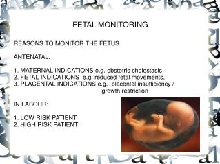
Why? • Two thirds of fetal deaths occur before the onset of labor. • Many antepartum deaths occur in women at risk for uteroplacental insufficiency. • Ideal test: allows intervention before fetal death or damage from asphyxia. • Preferable: treat disease process and allow fetus to go to term.
Antenatal monitoring • Uteroplacental insufficiency • Inadequate delivery of nutritive or respiratory substances to appropriate fetal tissues. • Inadequate exchange within the placenta due to decreased blood flow, decreased surface area or increased membrane thickness. • Inadequate maternal delivery of nutrients or oxygen to the placenta or to problems of inadequate fetal uptake.
Antenatal monitoring • Theoretical scheme of fetal deterioration • Fetal well being • Fetal growth retardation (Marginal placental respiratory function) • Fetal hypoxia with stress (Decreasing respiratory function) • Some residual effects of intermittent hypoxia (profound respiratory compromise) • Asphyxia • Death
Who? • Conditions placing the fetus at risk for UPI • Preeclampsia, chronic hypertension, • Collagen vascular disease, diabetes mellitus, renal disease, • Fetal or maternal anemia, blood group sensitization, • Hyperthyroidism, thrombophilia, cyanotic heart disease, • Postdate pregnancy, • Fetal growth restriction
How? • Methods for antepartum fetal assessment • Fetal movement counting • Assessment of uterine growth • Antepartum fetal heart rate testing • Biophysical profile • Doppler velocimetry
Fetal movement counting • Maternal perception of a decrease in fetal movements / change in the pattern of fetal movements may be a sign of impending fetal compromise. • Cardiff “count to ten” : 10 movements in 12 hours. • Kick charts • No robust evidence.
Reduced fetal movements • First episode: monitoring by CTG • Persistent: USS for growth, LV & UAD, CTG 2 to 3 times per week • Absent : • No FH: confirm by USS • FH present: growth scan and UAD
Tests for RFM • Cochrane data for CTG • Increased hospitalisation, additional tests & elective delivery • No benefit in outcome • Interpretation errors • False reassurance • UAD has been shown to be predictive of perinatal compromise in high risk pregnancies
Symphysiofundal height • General rule: fundal height in centimeters will equal the weeks of gestation. • Exceptions: maternal obesity, multiple gestation, polyhydramnios, abnormal fetal lie, oligohydramnios, low fetal station, and fetal growth restriction. • Customized chart :Abnormalities of fundal height should lead to further investigation. • Accuracy: poor?
Antenatal CTG • Initial observational studies showed a strong correlation between the abnormal CTG and poor fetal outcome • Widely used as the primary method of antenatal fetal assessment • Poor predictive value • High inter-observer inconsistencies
Antenatal CTG • Healthy fetuses display normal oscillations and fluctuations of the baseline FHR (Hammacher, 1966; Kubli, 1969). • Absence of these patterns was associated with increase in neonatal depression and perinatal mortality. • Accelerations of the FHR during stress testing correlated with fetal well being (Trierweiler, 1976).
Antenatal CTG • Accelerations of the FHR occur with fetal movement, uterine contractions, or in response to external stimuli. • FHR accelerations appear to be a reflection of CNS alertness and activity. • Absence of FHR accelerations seems to depict CNS depression caused by hypoxia, drugs, fetal sleep, or congenital anomalies.
CTG monitor • Cochrane: • Antenatal CTG has no significant effect on perinatal outcome or interventions such as early elective delivery • NICE: • Evidence does not support the routine use of antenatal electronic fetal heart rate monitoring for fetal assessment in women with an uncomplicated pregnancy and therefore it should not be offered
Decelerations • Decelerations- transient slowing of FHR below the baseline level of more than 15 bpm and lasting for 15 s or more
Antenatal CTG • Perinatal mortality: 6.2/1000 • False positive rate: 50% • False negative rate: 3.2 / 1000
Ultrasound • Quick, non-invasive procedure, easy interpretation • Customised fetal growth charts (serial scans) • Liquor volume • Placental function • Doppler study • Abnormal results correlate with increased risk of stillbirth and neonatal morbidity in selected pregnancies
Biophysical profile • Described by Manning (1980) • The number of biophysical activities that could be recorded increased with real time ultrasound: • Fetal movement (FM) • Fetal tone (FT) • Fetal breathing movements (FB) • Amniotic fluid volume (AFV)
Variables measured • CTG: reactive – as described earlier. • FBM: present - at least 1 episode of at least 30 seconds duration (within a 30 minute period). • FM: present - at least 3 discrete episodes. • FT: normal - at least 1 episode of extension of extremities or spine with return to flexion. • AFV: normal – largest pocket of fluid greater than 1 cm in vertical diameter.
Scoring • Biophysical profile (BPP) • Each variable • When normal: 2 • When abnormal: 0 • Highest Score: 10, Lowest Score: 0 • Accuracy improved by increasing the number of variables assessed. • Overall false negative rate: 0.6/1000
BPP and Perinatal morbidity • Significant inverse linear correlation (Manning, 1990) • Fetal distress • NICU admission • IUGR • 5 min Apgar <7 • Cord artery pH <7.20
Cochrane • Not enough evidence to evaluate the use of biophysical profile as a test of fetal well-being in high risk pregnancies except diabetes • No evidence of any benefit in screening • Errors associated with the BPP • Management decisions based on the score only. • Intervention based on a false positive low score • No intervention based on a false negative normal score • Management based on BPP without considering overall clinical findings.
Doppler velocimetry • 40% of combined ventricular output is directed to the placenta by umbilical arteries. • Assessment of umbilical blood flow provides information on blood perfusion of the fetoplacental unit. • Volume of flow increases and vascular impedance decreases with advancing Doppler velocimetry of the umbilical arteries gestational age. • Low vascular impedance allows a continuous forward blood flow throughout the cardiac cycle.
Doppler study • Doppler velocimetry • An increase in the vascular resistance of the fetoplacental unit leads to a decrease in end diastolic flow velocity or its absence in the flow velocity waveform. • Abnormal waveforms reflect the presence of a structural placental lesion. • Abnormal Doppler results require specific management protocols and intensive fetal surveillance.
Doppler velocimetry • Uterine arteries – 24/40
Doppler velocimetry • Umbilical arteries • Middle cerebral artery • Ductus venosus
Doppler study • Doppler velocimetry • A poor indicator of fetal compromise or adaptation to the placental abnormality but does identify patients at risk for increased perinatal mortality. • Strong association between high systolic to diastolic ratios and IUGR.
In summary Why? Who? How?