Download
1 / 56
640 likes | 2.76k Views
Intrapartum Fetal Monitoring. Partograph and Fetal Cardiotocography Prof. Azza Alyamani Department of Obstetrics &Gynecology. Aims of the presentation : 1. Be able to interpret routine data collected in labor. 2. Be confident to interpret a partogram and

E N D
Intrapartum Fetal Monitoring Partograph and Fetal Cardiotocography Prof. Azza Alyamani Department of Obstetrics &Gynecology
Aims of the presentation : 1. Be able to interpret routine data collected in labor. 2. Be confident to interpret a partogram and formulate a management plan based on the patterns observed. 3. Understand fetal response in labor. 4. Be able to interpret fetal monitoring . 5. Be familial of normal and abnormal fetal CTG traces.
Partogram Definition: it is graphical record of key data of labor progress with both maternal and fetal data entered against time . it is the process by which normal and abnormal progress of labor and also fetal response in labor can be defined.
Importance : It allows an instant visual assessment of the rate Cervical dilatation and comparison with an expected Norm , so that slow progress can be recognized Earl and appropriate actions taken to correct it Where possible.
Components Part 1 : Fetal condition a. Fetal heart rate. b. The condition of the membranes and liquor amnonii. c. Moulding and caput formation.
Part 2 : Progress of labor ( Cervicogram ): * it is a graphic representation of cervical dilatation and descent of the presenting part . * it is an essential part of the partogram . * it offer the chance of early detection of slow progress of labor. * first ,we set an alert line at 1cm/h. for the active phase dilatation to represent the ideal progress. then ,we set an action line 2-4h.later to the right of the alert line.
if the marks (plots) of progress falls beyond the action line , the progress of labor is slow and the cause of this should be sought and corrected if possible or the decision of cesarean section is considered.
Friedman′s Division of Labor : he divided the first stage of labor into: latent phase and active phases. then , he divided the active phase further into 3 parts : * acceleration phase. * phase of maximum slope and * deceleration phase.
Uterine contractions : Efficient powers ( good uterine contractions) are at the center of Efficient labor bec: * it cause cervical dilatation in the 1st stage , and * rotation & descent in the 2nd stage. efficient uterine contractions should be : 3-4 contractions in the 10 minutes ,each lasts 45-60 sec with amplitute of at least 50 mmHg.
Part 3 : Maternal condition Assessment of maternal condition regularly by monitoring : * vital signs; temperature , pulse and bl.P * drugs ; oxytocin ,analgesics ,IV fluids ect.. * investigations as urine analysis for protein and acetone and volume.
Management of labor using the partogram
Normal Progress of Labor *latent phase : 8 hours or less . *active phase : progress of the cervical dilatation remains on the alert line or between the alert and the action lines ( 1cm/hour ). small doses of oxytocin or/and ARM may be needed for active management of labor. *second stage : reseanable rotation and descent of the presenting part within 1 h. or less .
Abnormal (poor) Progress of Labor a. Prolonged Latent Phase: if extends 12 h (20h), in PG or 8h (14), in MG. b. Primary dysfunctional ( prolonged ) labor : slow cervical dilatation or poor descent . c. secondary arrest ( obstructed ) labor: of cervical dilatation in the 1st stage usually in the deceleration phase or arrest of the descent of the presenting part in the 2nd stage.
1= Prolonged latent phase. 2= primary dysfunctional labor ( protracted cervical dilatation). 3= primary dysfunctional labor ( protracted descent ). 5= secondary arrest of cervical dilatation. 6= secondary arrest of descent.
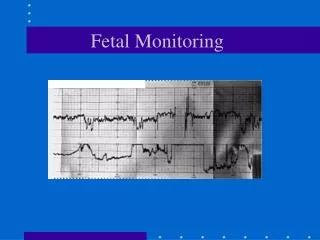
ElectronicFetal Heart Rate Monitoring (EFM)
Electronic fetal heart monitoring {EFM}: developed in the 1960s. is achieved by either : * internal or direct monitoring , by applying a bipolar electrode to the skin of the fetal scalp ,the cervix has to be dilated and membranes ruptured. * external or indirect monitoring. by using ultrasound ,usually by ; External toco dynamo metry with a pressure transducer placed on the uterus.
Indications for EFM *low risk women with; normal RHR on auscultation , no meconium staining liquor and normal progress of labor are extremely unlikely to deliver an asphyxiated infants. Admission test : initial 20 minutes EFM screen in early labor is enough to predict the likelihood of subsequent fetal hypoxia.
* high risk women; Maternal disorders: • hypertensive diseases. • diabetes mellitus. • renal diseases. • cardiac diseases. • APH. • RH isoimmunization.
Labor complications : • dysfunctional labor ( slow progress). • oxytocin augmentation or induction . • preterm labor. • VBAC. Fetal complications : • IUGR. • previous SB. • meconium staining. • Post term pregnancy. • abnormal FHR on auscultation. • twins.
Fetal Heart Rate Patterns A. Baseline FHR : * tachycardia. * bradycardia. * variability. B. Periodic FHR : * Accelerations. * decelerations. • early • prolonged • variable • mixed • late • sinusoidal
Baseline FHR: * It is the rate recorded in between uterine contractions. * Normally 110 – 150 bpm. * usually assessed over 15 minute interval during labor.
Baseline tachycardia * It is a FHR above 150 - 160 bpm. * causes : • Fetal hypoxia. • Maternal or Fetal infection. • Drugs as ritodrine and atropine. • Maternal hyperthyrodism. • maternal dehydration. • Fetal anemia . • Fetal cardiac arhythmias.
Baseline bradycardia * It is a FHR less than 90 -110 bpm. * It may be mild ( 90 -110 bpm. ) or severe ( < 90 bpm. ). * persistent mild fetal bradycardia is usually benign. While severe bradycardia caused by fetal congenital heart block.
Variability * baseline or beat - to -beat variability is controlled by the balance between the sympathetic and parasympathetic nervous control of the heart. * normal variability is 5 – 25 bpm. * it is reduced < 5 bpm. in : • fetal hypoxia. • physiological during fetal sleep cycles. • drugs as ;dethadine ,barbiturates and atropine.
Periodic FHR periodic changes in the FHR may be either : A. Accelerations : * is associated with ; • fetal movements. • uterine contractions. * are considered benign and its presence indicates a well oxygenated fetus.
normal FHR accelerations associated with fetal movements ( reactive NST )
normal FHR accelerations with uterine contractions
B. decelerations : classically 3 well defined decelerations are described ; early ,variable and late. 1. Early deceleration : • this deceleration have a uniform shape ( bell), starts early in the contraction and mirrors it. • the magnitude of the deceleration is <40bpm. • cause : head compression mediated by vagal reflex. • it occurs during the active phase , they are benign .
2.Variable deceleration : • it is the most common deceleration pattern. • it appears as abrupt fall and return in FHR, preceded and followed by small accelerations ( shoulders). • they are variable in shape V ,U or W shape , duration and timing. • the magnitude is usually 50-80 bpm. • cause : cord compression. if it persists fetal hypoxia occurs. • mild variable deceleration ( last < 30 sec.) is benign. moderate (last 30 -60 sec.) and severe (last > 60 sec.) indicates fetal hypoxia.
3. Late deceleration: • it have a similar shape and magnitude as early deceleration but their timing is different. • it start as the contraction peaks and does not return to the baseline FHR until after the end of the contraction. • cause : fetal hypoxia.
Interpretation of FHR Patterns First identify : 1. baseline FHR. 2. presence , reduced or loss of variability. 3. presence of accelerations or decelerations. 4. if there is decelerations ,its frequency ,type and severity .
Types of FHR patterns: a. reassuring or normalpattern. * Baseline fetal heart rate between 110 and 150 bpm. * Good variability. * Presence of accelerations . * Absence of significant decelerations.