Download
1 / 45
512 likes | 1.29k Views
Fetal Monitoring & Wellbeing. Fetal Well-being. Principles: the ideal scheme to assess FWB should: Take account of cycles of normal fetal behavior detect impending harm accurately and in time to intervene to prevent it give reassurance preferably up to 7 days

E N D
Fetal Monitoring & Wellbeing
Fetal Well-being • Principles: • the ideal scheme to assess FWB should: • Take account of cycles of normal fetal behavior • detect impending harm accurately and in time to intervene to prevent it • give reassurance preferably up to 7 days • avoid causing unnecessary anxiety • allow detection of specific causes e.g hypoxia, infection, malf’n • produce measurable benefits in reducing perinatal loss/injury • such system is likely to involve tests which assess several fetal systems, CVS, NS,, RS and use >1 modality
Fetal Monitoring- The Evidence ? Factors for increased fetal risk • Medical complications: • HTN, DM, AID, Hb pathies • Fetal problems: • IUGR, Non-lethal anamolies, prematurity, postdatism, hydrops • IU problems: • Bleeding, fever, meconium stain, oxytocin augment.
Fetal Monitoring- The Evidence ? Utero-placental complex • Uterus depends on placenta for diffusion of nutrients and respiratory gas exchange. • Placental function depends on uterine blood flow (UBF) • Uterine contraction leads to transient decreased UBF • Borderline placenta may lead to fetal asphyxia during L&D • Fetal compensatory responses limit the damage • Prolonged or severe hypoxia may cause injury or death.
Fetal Well-being • Invasive: • Chorion villus Sampling • Amniocentesis • Umbilical artery canulation Non-invasive: • Fetal Movement Count: • Fetal Heart Recording….. CTG • Biophysical Profile {BPP} scoring • Doppler studies
Fetal Monitoring- The Evidence ? Intrapartum Fetal Assessment • Electronic Fetal Heart Monitoring • Fetal Scalp pH ( and pCo2, pO2) Monitoring • Fetal Scalp Stimulation • Vibroacoustic Stimulation • UA Velocimetry and Biophysical profile • Fetal Pulse Oximetry • Near-infrared Spectroscopy
C ardio- T oco- G raph Screening tool to assess the fetal state of oxygenation and predicts early signs of hypoxia and fetal distress.
Cardiotocography Components • Stimulus: Contractions/ fetal movements • Baseline fetal heart rate • Baseline variability • Accelerations • Decelerations
Cardiotocography How to read a CTG • baseline heart rate
Cardiotocography How to read a CTG • Baseline variability • Classification: • Silent 0-5 bpm • Reduced 6-10 bpm • Normal 11-25 bpm • Saltatory >25 bpm
Cardiotocography • accelerations
Cardiotocography • Decelerations:
Cardiotocography • Decelerations:
Basal fetal oxygenation. The relationship of late decelerations to baseline fetal oxygenation during contractions
How to Read CTG Patient's data + Date & Time DR C BRAVADO Signature + Date & Time
How to Read CTG DR C BRAVADO VARIABILITY ACCELERAT’N DECELERAT’N CONTRACTIONS DEFINE RISK BASELINE RATE OVERALLASSESSMENT
classification of Fetal Heart Rate Pattern Normal Pattern • Baseline Rate 110-150 bpm • Amplitude of baseline variability 5/10-25 bpm • Absence of decelerations, except for fleeting& short • Presence of 2 or more accelerations during a 20 min period
classification of Fetal Heart Rate Pattern Suspicious pattern • Baseline rate of 150-170 bpm/ 100-110 bpm • Amplitude of variability bn 5-10 bpm > 40 min • Increased variability above 25 bpm {saltatory} • Absence of accelerations for > 40 min • Sporadic decelerations of any type, unless severe
classification of Fetal Heart Rate Pattern Any of the following: Pathological Pattern • Baseline heart rate < 100 bpm or > 170 bpm • Variability < 5 bpm for > 40 min • Recurrent decelerations of any type • Severe variable or late decelerations • A sinusoidal pattern
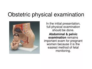
Ultra Sound Scan • Indications of use: • pregnancy location • viability • fetal number • dating • anomaly • placental localization, amniotic fluid • fetal growth and wellbeing • during invasive procedures
Assessment of fetal state • Assessment of gestational age and fetal growth: • menstrual history unreliable in up to 45% of women • serial fundal height measurement provides a guide to fetal growth • USS: crown-rump length before 14 weeks • USS: BPD serial measurement every 2 weeks for fetal growth. Unreliable after 28 weeks for dating • USS: head/abd ratio, 2 weeks serial HC & AC for fetal growth .. IUGR AC< but initially HC ~. • USS: femur length, more precise guide to gestational age than BPD
Serial measurements are necessary to identify the growth pattern and detect any lag in the growth and IUGR
Biophysical Profile& Color Doppler ultrasound in the high risk pregnancy
BPP is applying to detect prenatal asphyxia • Doppler ultrasound is a modality for detecting fetal hypoxia and acidosis • Doppler can also predict later pre- eclampsia at the 24-26 gestational weeks.
Hypoxia: Low Oxygen tension • Asphyxia: Low Oxygen and high CO2 • Ischemia: Drop in blood flow
Biophysical Profile • BPP uses FHR monitor and real time USS to assess: • fetal breathing movement • discrete body or limb movement • fetal tone • FHR • amniotic fluid volume • Amniotic fluid volume is most important • Fetal breathing movement is the first to disappear in asphyxia • 7 days reassurance in low risk, only 24 hours in high risk preg
comment • As you know, oligohydramnios may be • Mild AFI=5-8cm • Moderate AFI=2-5cm • Sever AFI<2cm only sever oligohydramnios is considered as an abnormal score.
Fetal movement and fetal tone develop between 7.5 and 9 weeks’ menstrual age • Fetal breathing movements are detectable by, at least 17-18 weeks’ gestation • The non-stress test is most reliable between 32 weeks and term (Ware, 1994).
l An early stage in fetal adaptation to hypoxemia - central redistribution of blood flow ( brain-sparing reflex) • increased blood flow to protect the brain, heart, and adrenals • reduced flow to the peripheral and placental circulations
Clinical Indications for Doppler Studies • most useful in assessing IUGR • identify only the sub-group which is hypoxemic bec/of inadequate placental function and may be abnormal for up to 18 weeks before any fetal problem is observed • no proven role in population screening for increased risk of pre-eclampsia or IUGR
Practice read the following CTGs
Practice Plot the growth chart