Download
1 / 67
690 likes | 1.1k Views
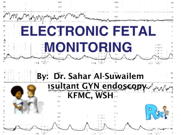
Electronic Fetal Monitoring. Terri Imus, RN. Electronic Fetal Monitoring. Indications for continuous EFM Any pregnancy considered high risk Induction or augmentation of labor Decreased fetal movement Premature labor Premature rupture of membranes. Oligohydramnios Hypertension

E N D
Electronic Fetal Monitoring Terri Imus, RN
Electronic Fetal Monitoring Indications for continuous EFM Any pregnancy considered high risk • Induction or augmentation of labor • Decreased fetal movement • Premature labor • Premature rupture of membranes
Oligohydramnios • Hypertension • Abnormal fetal heart rate • Fetal malpresentation in labor • IDDM • Multiple Gestation • Previous C/S • Trauma • Meconium
ACOG & AAP • When EFM is the method selected for fetal assessment. The MD & obstetrical personnel should be qualified to identify and interpret abnormalities. These guidelines also state that it is appropriate for MD & Nurse to use the descriptive terms that have been given to fetal monitoring patterns in charting and reporting • Those not qualified or are unsure of the interpretation in FHR patterns should seek other professionals to assist in this evaluation and interpretations • The nurse should document the presence of MD and nurse, pt position and changes in cervix,
Therapeutic interventions such as O2 and medications • Increased or decreased BP • Febrile • Amniotomy, AROM,SROM, color amt. consistency • Is the patient complete/pushing • All of these descriptive details give a picture that indicates what is going on with the patient and possible cause of change in FHR pattern
Guidelines emphasize that when there is a change in the FHR pattern all of those things should be documented as well as a return to baseline • Each tracing should include • Pt Name • ID # • Date, Time of admission/delivery • EDC, Gravida Para and any other identifying information • AAP/ACOG
ACOG • Has not identified core competencies in FHR monitoring Standard guidelines Norm 110-160 • Fetal tachycardia • Mod 161-180 • Marked “ 181-more • Fetal Bradycardia • Mod 100-119 • Marked” 90 or less
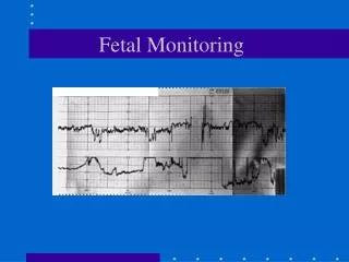
4 Basic Features • Baseline • Variability • Bradycardia <110 bpm • Tachycardia >160 bpm Periodic changes: FHR accelerations or decelerations that occur with contractions. Decelerations are routinely described as early, late, or variable.
Non-periodic changes (no changes in variability) Nonperiodic changes can occur spontaneously, without contraction activity, and are also described as accelerations or decelerations. Variable decelerations can appear during a Non-stress test and may be a sign of cord compression or oligohydramnios, both of which can have adverse effects on the fetus.
Baseline Variability • Normal FHR 5 bpm greater than or equal to 5 bpm, between contractions Nonreassuring FHR less than 5 bpm or less, but less than 30 min of tracing Abnormal FHR less than 5 bpm for 90 min or more.
Baseline variability • The minor fluctuations on baseline FHR at 3-5 cycles p/m will reflect baseline variability • Examine 1 min segment and estimate highest peak and lowest trough • Normal is more than or equal to 5 bpm
Factors affecting Baseline variability • Para-Sympathetic affects short term variability • Sympathetic affects long term • CNS Drugs reduces Variability
Increased gestational age may increase variability • Mild Hypoxia may cause both Sympathetic and Parasympathetic stimulation
Accelerations • Accelerations transient increase in FHR of 15 bpm or more lasting for 15 sec • Absence of accelerations on an otherwise normal Fetal heart tracing remains unclear • Presence of FHR Accelerations usually have good outcome
Early Decelerations • Head compression Begins on the onset of contraction and returns to baseline as the contraction ends Should not be disregarded if it appears early in labor or in the antenatal period
Late Decelerations • Uniform periodic slowing of FHR with the on set of the contraction Reduced baseline variability together with late decelerations and repetitive late deceleration increases risk of fetal acidosis and an Apgar score of less than 7 at 5/min with an increased risk of adverse outcome
Late Decelerations Due to acute and chronic utero-placental insufficiency • Occurs after the peak and past the length of uterine contraction, often with slow return to the baseline • Is precipitated by hypoxemia • Associated with respiratory and metabolic acidosis • Common in patients with PIH, DM, IUGR or other forms of placental insufficiency
Variable Decelerations • Variable intermittent periodic slowing of FHR with rapid onset recovery and isolation • They can resemble other types of deceleration in timing and shape • Atypical associated with an increased risk of umbilical artery acidosis and Apgar score less than 7 at 5 min
Additional components Loss of 1 or 2 degree rise in baseline rate • Slow return to baseline FHR after and end of contraction • Prolonged secondary rise in Base FHR • Biphasic deceleration • Loss of variability during deceleration • Continuation of the baseline at a lower rate
Variable Deceleration (Vagal activity) Inconsistent in configuration • No uniform temporal r-ship to the onset of contraction, are variable and occur in isolation • Worrisome when Rule of 60 is exceeded (i.e. decrease of 60 bpm,or rate of 60 bpm and longer than 60 sec)
Caused by compression of the umbilical cord • Often associated with Oligo-hydramnios with or without rupture of membranes • Acidosis if prolonged and recurrent
Prolonged Deceleration Drop in FHR of 30 bpm or more lasting for at least 2 mins • Is pathological when it crosses 2 contractions in 3 mins • Results in reduced of O2 transfer to placenta • Associated with poor neonatal outcome
Prolonged DecelerationsCAUSES • Cord prolapse • Maternal hypertension/hypotension • Uterine hypertonia • Epidural/spinal or pudendal anesthesia • Can follow a vag exam, AROM or SROM with high presenting part
Intrauterine Resuscitation Have the mother lie on her left/right side or in a knee chest position To alleviate possible cord compression Reduce or stop any oxytocin • Initiate tocolysis • To decrease uterine activity and increase placental blood flow • Increase IV fluid • To increase maternal blood/fluid volume Give oxygen @ 10-12 L/min via mask
Physician may apply an internal monitorto verify the accuracy of external monitor reading • Physician may administer amnioinfusion to decrease pressure on cord or dilute mec. • If the heart rate is not restored to normal within 30 minutes, prompt delivery is needed. Cesarean section may then become necessary. Goal is to deliver ASAP
Causes of Baseline Change • Postdates • Drugs • Idiopathic • Arrhythmias • Hypothermia • Increased vagal tone • Cord Compression Management depends on the clinical situation
Causes of Bradycardia • Asphyxia • Drugs • Prematurity • Maternal Fever • Maternal thyrotoxicosis • Maternal Anxiety • Idiopathy Management depends on the clinical situation
Baseline Tachycardia • Asphyxia • Drugs • Prematurity • Maternal fever • Maternal thyrotoxicosis • Maternal Anxiety • Idiopathy
Sinusoidal Pattern • Regular Oscillation of the Baseline long-term Variability resembling a Sine wave fixed cycle of 3-5 p min with amplitude of 5-15bpm and above but not below the baseline • Should be viewed with suspicion as poor outcome has occurred (maternal/fetal hemorrhage)
Sinusoidal pattern - distinctive smooth undulating Sine-wave baseline Cord compression • Hypovolemia • Ascites • Idiopathic (fetal thumb sucking) • Analgesics • Anemia • Abruption Management depends on clinical situation
Summary of tracing • Normal with all 4 Features • Suspicious one non reassuring category and remainder are reassuring • Pathological 2 or more non-reassuring categories or one or more abnormal categories.
At Birth Need to Consider Cord pH if tracing suspicious Preterm labor Mec. stained amniotic fluid FBS intrapartum (lab availability) Lack of tone delivery Operative or instrumental delivery
COMMUNICATION • DESCRIBE THE PATTERN ACCURATELY • MAKE AN ATTEMPT TO ASSESS WHETHER THE FETUS IS IN TROUBLE • IF YOU WANT THE PHYSICIAN THERE, COMMUNICATE THAT • THE NURSE HAS MORE DATA THANTHE PHYSICIAN
Communication • SYSTEMATIC APPROACH REDUCES ERRORS • DESCRIBE WHAT YOU SEE • AVOID THE NEED TO CLASSIFY EVERY DECELERATION • ASSESS THE OVERALL CONDITION OF THE FETUS
Electronic Fetal Monitoring • Improve knowledge for all staff • Improve clinical skills • Training should include instruction on documentation and storage • Training should include appropriate clinical responses to suspicious or pathological tracings • Training should include local guidelines relating to fetal monitoring both intermittent and EFM
DOCUMENTATION OF COMMUNICATION • DO NOT JUST SAY THAT Dr. Whoduneit WAS NOTIFIED • RECORD THE PHYSICIAN’S RESPONSE and any ORDERS
COMMUNICATION • DESCRIBE FHR PATTERN • I AM CONCERNED ABOUT THE CONDITION OF THIS BABY • IT IS OMINOUS AND NON-REASSURING • IF PERSISTENT, REQUIRES PHYSICIAN EVALUATION
COMMUNICATION • THE FETUS HAS INCREASED VARIABILITY AND THE BASELINE IS HARD TO NTERPRET • PHYSICIAN PRESENCE NOT REQUIRED
COMMUNICATION • NOTIFY IF NO DRUGS WERE GIVEN • THE FETUS HAS HAD A SINUSOIDAL PATTERN FOR 20 MINUTES. I HAVE NOT GIVEN ANY NARCOTICS AND THE PATTERN PERSISTS DESPITE POSITIONING, HYDRATION AND OXYGEN. • PHYSICIAN PRESENCE MAY NOT BE REQUIRED but inform
COMMUNICATION what if • THE FETUS SUSTAINED A PROLONGED DECELERATION ASSOCIATED WITH HYPERSTIMULATION • THE PATTERN RESOLVED AFTER …. • PHYSICIAN PRESENCE MAY NOT BE IMMEDIATELY REQUIRED, BUT SHOULD BE NOTIFIED