Bone Formation and Types: An Overview
Learn about bone formation, types of bones, and the function of bones. Discover how bone grows and changes throughout life, and the composition of bone matrix. Explore the two types of bone formation and the three types of bone cells. Understand the classification of bones and the identifying characteristics of bones. Gain knowledge about the skeletal system, including the axial and appendicular skeleton, as well as the structure of the skull.

Bone Formation and Types: An Overview
E N D
Presentation Transcript
Bone Fractures Bone Formation Facial Bones Types of Bones Osteology Arthrology Skeletal System Skull Appendicular Skeleton Axial Skeleton Function of Bone Cartilage Classification of Bones Bone Disorders
Osteology – The Science of Bones Bone is living tissue it grows and changes throughout your life. You completely replace your entire skeleton about every ten years. Bone are composed of: Cells, Fibers and a Matrix ( Ca2+) The Matrix of the Bone is referred to as an Hydroxyapatite The Chemical Composition of the bone matrix is (Ca2+)10(PO4)6 (OH)2 return
Figure 07.01 Structure of a Long Bone Return
Bone Formation Two Types - • Intramembranous – Develops in the connective tissue membranes in the embryo called Mesenchyma – embryonic CT • Endochrondral Bone Formation – Bone forms in cartilage A cartilage model of the skeleton is in place and the cartilage is removed and bone is laid down. NOTE : Bone is NOT calcified cartilage Return
Formation of bone is called Ossification - it starts in the embryo and continues to age 18 in females and 20 in males. Within the cartilage skeleton model of the embryo are ossification centers. This is where the cartilage is being destroyed and the bone is being made. Ossification takes place in the middle of the bone shaft first and then at the ends. The bone develops moving very slowly toward the ends of the bone. Return
Two types of bone are produced : Spongy Bone – Contains a lot of cavities this structure reduces weight and increases strength of the bone. This is the region that is effected by osteoporosis the most. Hemopoietic tissue (makes blood) fills the cavities of spongy bone. Compact Bone - Dense strong bone that makes up the outer surface of the bone. Compact bone is composed of millions of subunits called: Haversian Systems or Osteons Return
Figure 07.02 Return
Diaphysis Volksman Canal Epiphyseal Plate or Growth Plate Articular Cartilage Spongy Bone with Hemopoietic Tissue Return Epiphysis
Three types of Bone Cells • Osteoblasts – Makes new bone • Osteocytes – Maintain existing bone if damage occurs. Live inside calcified bone in lacuna (“little pond” in Latin) • Osteoclasts – Remove bone, act like white blood cells. They are triggered by parathormone from the thyroid to breakdown bone to increase calcium level in blood. Their action can lead to osteoporosis – Loss of bone mass due to lack of calcium. Bones can become weak and brittle. Return
Haversian System or Osteon Lamella of bone Canaliculi Haversian Canal Osteocyte Haversian Canal Systems Return
Function of Bones • Support • Motion – Muscle Attachment • Protection – Skull protects Brain Ribs & Sternum protects Heart & Lungs 4. Calcium Storage – There is a constant exchange between bones and blood. 5. Bone Marrow – Process called Hemopoisis is the making of Red and White blood cells. In the infant red marrow is found in most of the bones. In the adult it is only found in 1. Sternum and Ribs 2. Hip Bones (iliac Crest) Take marrow samples here 3. Bodies of Vertebrae 4. Proximal end of Long Bones return
Classification of Bones • Long Bones Upper Extremities - Humerus Radius & Ulna Lateral Medial Palm - Metacarpals Fingers - Phalanges Lower Extremities - Femur Tibia & Fibula Medial Lateral Foot - Metatarsals and Phalanges return
2. Short Bones - Wrist - Carpal bones or carpus Ankle - Tarsals bones or Tarsus 3. Irregular Bones - Vertebrae & Skull may be movable or immovable contain many irregular bones 4. Flat Bones - Scapula – Shoulder blade Clavicle – Collar bone Skull - Frontal & Parietal bones of skull 5. Sesamoid Bones “seed” – small, nodular found within tendon ex: patella (knee cap) return
Identifying Characteristics of Bones 1. Fossa – Depression in a bone ex: TMJ (Temporal Mandibular Joint) forms a fossa 2. Sinus – Cavity in a bone ex: maxillary sinus – cavity above upper jaw 3. Foramen – Hole in a bone ex: Foramen Magnum – hole for spinal cord in skull 4. Meatus – Tubular structure in bone ex: External auditory meatus let sound enter skull 5. Condyles – Large smooth curved surfaces that touches another bone. ex: Distal end of femur 6. Trochanter – A large projection on a bone for muscle attachment Lec 1 return
Figure 07.09 Axial Skeleton Appendicular Skeleton Return
Skeleton Axial Skeleton - Skull Ossicles (ear bones) Hyoid Sternum & Ribs Vertebral column Appendicular Skeleton - Upper Extremities Pectoral Girdle - clavicle & scapula (attachment to axial skeleton) Lower Extremities Pelvic Girdle – ilium, ischium, pubic bone Return
Skull Two major components – • Skull Cap - Calvarium or cranium contains 8 bones that enclose the brain. • Facial Bones – 14 bones that support muscles of the face. Bones of the Calvarium – 1. Frontal – forehead – Anterior fossa of base of skull 2. Parietal – 2 – means “walls”- top of skull 3. Temporal – 2 – sides of skull 4. Occipital – posterior part of skull 5. Sphenoid 6. Ethmoid 1 2 5 6 3 4 Return
Temporal Bone - Four Parts • Squamous – Flat/thin part of skull ( Don’t get hit here) • Mastoid Process – “breast” – contain sinus for middle ear. • Zygomatic Process – “bar” – posterior portion of cheek bone • Petrous portion – “hard” – houses the inner ear Occipital Bone – Thick Bone Contains the Foramen Magnum “Big Hole” Articulates with first cervical vertebra - Atlas Greek god Atlas Return
Sphenoid – Floor of skull – called the keystone of the skull keeps the other bones in place. Holds the Pituitary Gland in the “Turkish Saddle” formed by the four Clinoid processes. Sphenoid Ethmoid Ethmoid Bone – “sieve” contains holes for olfactory nerves to pass through. This is called the Cribriform Plate. Return
Looking down into skull Pituitary would be here Clinoid Processes “Turkish Saddle” Squamous Petrous portion V shape of Occipital is called a Lambdoidal Shape Return
The immovable joints between the bones of the skull are called Sutures Anterior Return Posterior
Infant Skull – Contains areas of connective tissue called Fontanelles Allows the skull to move during birth and accommodates rapid growth of brain. Return
3 4 Facial Bones There aretwo of each of the facial bones 1. Maxilla – Upper Jaw called keystone of face all facial bone touch the maxilla except mandible. 2. Zygomatic Process – Anterior roof of mouth formed by maxilla 3. Nasal Bones – Forms bridge of nose 4. Lacrimal Bones – Inferior medial orbit 5. Zygomatic Bones – Middle of cheek 6. Palatine Bones – Posterior portion of the roof of the mouth 7. Vomer – Bone of lower septum of nasal cavity 2 5 7 1 Return
The Cheek is made up of : Zygomatic Process - Maxilla Zygomatic Bone Zygomatic Process - Temporal The roof of the mouth is called the Hard Palate It is composed of the: Maxilla - Anterior Palatine - Posterior Return
Nose – Lateral Nose - Two inferior Conchae in the Inferior lateral nasal cavity Medial Nose - Vomer – medial nasal cavity (called the Septum) Mandible – Jaw bone only moveable joint of skull Coronoid Process Condyloid Process Part of TM Joint Ramus “branch” Angle Body Return
Figure 07.12 Return
Teeth—Humans have a heterodont dentition ( Different Teeth)
Figure 07.17 C1 C7 T1 Ribs attach to all the Thoracic Vertebrae T12 L1 L5
C1 – Atlas C2 – Axis Odontoid Process “tooth” connects these two together allows you to rotate your head Figure 07.18 Laminectomy – Cut through this part of the vertebra to get to the spinal cord Muscle attachment Spinal cord found here Longest spinous processes in Thoracic region Rib attachment Thoracic is area most common for tumors Largest bodies in Lumbar region due to carrying all of the body’s weight
Vertebral Column – Posterior Lateral Anterior Cervical Thoracic Scoliosis Lumbar Sacral Abnormal Curves – Kyphosis – Hunchback – Accentuated Thoracic Curve Lordosis – Swayback – Accentuated Lumbar Curve Scoliosis – Lateral curve in Thoracic region more common in females begins during puberty. Coccyx
Intervertebral Discs – Rings of Fibrocartilage between the vertebrae Annulus Fibrosis Stiff cartilage ring Nucleus Pulposus – Soft gelatinous center Slipped Disc - When someone has a slipped disc the disc doesn’t really move. The Annulus tears and the soft gelatinous center gets squeezed out. Sort of like putting pressure on a jelly donut the jelly squeezes out. Best cure is bed rest and to let the ring heal and the center to regenerate.
Figure 07.21 Return
Deltoid Tuberosity Surgical Neck Humerus – Bone of Arm
Figure 07.25 Proximal Semilunar notch Lateral Medial Distal
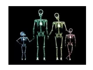
Figure 07.29 Female Male
Figure 07.30 Common fracture site Femur For muscle attachment, you can see how active person is by looking at this area of a bone. Patella in tendon found here
Figure 07.31 Articulates with femur Articulates with Talus Bumps on side of ankle
Figure 07.33 Dancer’s Fracture Return
Bone Fractures Box Figure 07.01a