Download
1 / 39
390 likes | 404 Views
Explore the application of genetic analysis and DNA technology in the study of development, using model organisms like fruit flies, nematodes, and mice. Learn about the processes of cell division, differentiation, and morphogenesis in both animals and plants. Discover how differential gene expression leads to the specialization of cells in a multicellular organism. Explore the concept of totipotency in plants and the cloning of organisms through nuclear transplantation.

E N D
Chapter 21 • The Genetic Basis of Development
From Single Cell to Multicellular Organism • Application of genetic analysis and DNA technology has revolutionized study of development
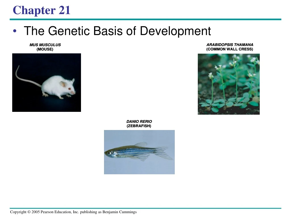
Figure 21.1 • Researchers • Use mutations to deduce developmental pathways • Use tools of molecular genetics developmental biology
DROSOPHILA MELANOGASTER (FRUIT FLY) CAENORHABDITIS ELEGANS (NEMATODE) 0.25 mm • Model organisms Figure 21.2
ARABIDOPSIS THAMANA (COMMON WALL CRESS) MUS MUSCULUS (MOUSE) DANIO RERIO (ZEBRAFISH)
(b) Tadpole hatching from egg (a) Fertilized eggs of a frog Figure 21.3a, b • Zygote organism • cell division, cell differentiation, and morphogenesis
Mitosis • Zygote large # of cells • Cell differentiation • Specialization in structure and function • Morphogenesis • Shape to the organism and its parts
(a) Animal development. Most animals go through some variation of the blastula and gastrula stages. The blastula is a sphere of cells surrounding a fluid-filled cavity. The gastrula forms when a region of the blastula folds inward, creating a tube—a rudimentary gut. Once the animal is mature, differentiation occurs in only a limited way—for the replacement of damaged or lost cells. Gut Cell movement Zygote (fertilized egg) Eight cells Blastula (cross section) Gastrula (cross section) Adult animal (sea star) Cell division Morphogenesis Observable cell differentiation (b) Plant development. In plants with seeds, a complete embryo develops within the seed. Morphogenesis, which involves cell division and cell wall expansion rather than cell or tissue movement, occurs throughout the plant’s lifetime. Apical meristems (purple) continuously arise and develop into the various plant organs as the plant grows to an indeterminate size. Seed leaves Shoot apical meristem Root apical meristem Zygote (fertilized egg) Two cells Embryo inside seed Plant Figure 21.4a, b • 3 processes of development overlap in time
Different cell types result from differential gene expression in cells with the same DNA • Differences between cells in a multicellular organism f/ differences in gene expression
Totipotency in Plants • Differentiated cell generates a whole organism
Transverse section of carrot root CONCLUSION EXPERIMENT 2-mg fragments Fragments cul- tured in nutrient medium; stir- ring causes single cells to shear off into liquid. Single cells free in suspension begin to divide. Embryonic plant develops from a cultured single cell. Plantlet is cul- tured on agar medium. Later it is planted in soil. A single Somatic (nonreproductive) carrot cell developed into a mature carrot plant. The new plant was a genetic duplicate(clone) of the parent plant. RESULTS Adult plant At least some differentiated (somatic) cells in plants are toipotent, able to reverse their differentiation and then give rise to all the cell types in a mature plant. Figure 21.5
Cloning • One or more somatic cells from a multicellular organism to make another genetically identical individual
Nuclear Transplantation in Animals • Nucleus of an unfertilized egg cell or zygote is replaced with the nucleus of a differentiated cell
Researchers enucleated frog egg cells by exposing them to ultraviolet light, which destroyed the nucleus. Nuclei from cells of embryos up to the tadpole stage were transplanted into the enucleated egg cells. EXPERIMENT Frog tadpole Frog egg cell Frog embryo Fully differ- entiated (intestinal) cell Less differ- entiated cell Donor nucleus trans- planted Enucleated egg cell Donor nucleus trans- planted <2% develop into tadpoles Most develop into tadpoles Figure 21.6 • Experiments with frog embryos
RESULTS Most of the recipient eggs developed into tadpoles when the transplanted nuclei came from cells of an early embryo, which are relatively undifferentiated cells. But with nuclei from the fully differentiated intestinal cells of a tadpole, fewer than 2% of the eggs developed into normal tadpoles, and most of the embryos died at a much earlier developmental stage. CONCLUSION The nucleus from a differentiated frog cell can direct development of a tadpole. However, its ability to do so decreases as the donor cell becomes more differentiated, presumably because of changes in the nucleus.
Egg cell donor Mammary cell donor APPLICATION This method is used to produce cloned animals whose nuclear genes are identical to the donor animal supplying the nucleus. 1 2 Egg cell from ovary Nucleus removed Nucleus removed Cells fused Cultured mammary cells are semistarved, arresting the cell cycle and causing dedifferentiation 3 TECHNIQUE Shown here is the procedure used to produce Dolly, the first reported case of a mammal cloned using the nucleus of a differentiated cell. Nucleus from mammary cell Grown in culture 4 RESULTS The cloned animal is identical in appearance and genetic makeup to the donor animal supplying the nucleus, but differs from the egg cell donor and surrogate mother. Early embryo Implanted in uterus of a third sheep 5 Surrogate mother Embryonic development 6 Lamb (“Dolly”) genetically identicalto mammary cell donor Figure 21.7 Reproductive Cloning of Mammals (1997)
Figure 21.8 • “Copy Cat” • First cat cloned
Problems with Animal Cloning • Sm. % of cloned embryos develop normally • ‘old’ cells
The Stem Cells of Animals • Relatively unspecialized cell • Can reproduce itself indefinitely • Differentiates into specialized cells
Embryonic stem cells Adult stem cells Early human embryo at blastocyst stage (mammalian equiva- lent of blastula) From bone marrow in this example Totipotent cells Pluripotent cells Cultured stem cells Different culture conditions Liver cells Blood cells Nerve cells Different types of differentiated cells Figure 21.9 • Stem cells isolated from early embryos at the blastocyst stage
Adult stem cells • Pluripotent (able to give rise to multiple but not all cell types)
Cell determination • Precedes differentiation, expression of genes for tissue-specific proteins
2 1 Nucleus Master control gene myoD Other muscle-specific genes DNA OFF OFF Embryonicprecursor cell Determination. Signals from othercells lead to activation of a masterregulatory gene called myoD, andthe cell makes MyoD protein, atranscription factor. The cell, nowcalled a myoblast, is irreversiblycommitted to becoming a skeletalmuscle cell. OFF mRNA MyoD protein(transcriptionfactor) Myoblast (determined) Differentiation. MyoD protein stimulatesthe myoD gene further, and activatesgenes encoding other muscle-specifictranscription factors, which in turn activate genes for muscle proteins. MyoD also turns on genes that block the cell cycle, thus stopping cell division. The nondividing myoblasts fuse to become mature multinucleate muscle cells, alsocalled muscle fibers. mRNA mRNA mRNA mRNA Myosin, othermuscle proteins,and cell-cycleblocking proteins MyoD Anothertranscriptionfactor Muscle cell(fully differentiated) • Determination and differentiation of muscle cells Figure 21.10
Unfertilized egg cell Molecules of a a cytoplasmic determinant Fertilization Nucleus Zygote (fertilized egg) Mitotic cell division Two-celled embryo (a) Cytoplasmic determinants in the egg. The unfertilized egg cell has molecules in its cytoplasm, encoded by the mother’s genes, that influence development. Many of these cytoplasmic determinants, like the two shown here, are unevenly distributed in the egg. After fertilization and mitotic division, the cell nuclei of the embryo are exposed to different sets of cytoplasmic determinants and, as a result, express different genes. Figure 21.11a • Cytoplasmic determinants regulate expression of genes that affect developmental fate of cells Molecules of another cyto- plasmic deter- minant Sperm Sperm
Induction by nearby cells. The cells at the bottom of the early embryo depicted here are releasing chemicals that signal nearby cells to change their gene expression. (b) • Induction • Signal molecules from embryonic cells cause transcriptional changes in nearby target cells Early embryo (32 cells) Signal transduction pathway NUCLEUS Signal receptor Signal molecule (inducer) Figure 21.11b
Pattern formation • Development of a spatial organization of tissues and organs • Occurs continually in plants • Limited to embryos and juveniles in animals
Positional information • Molecular cues that tells a cell its location relative to other cells
Follicle cell Nucleus Egg cell developing within ovarian follicle Egg cell Nurse cell Fertilization Laying of egg Fertilized egg Egg shell Nucleus Embryo Multinucleate single cell 3 4 6 7 1 5 2 Early blastoderm Plasma membrane formation Yolk Late blastoderm Cells of embryo Body segments Segmented embryo 0.1 mm Hatching Larval stages (3) Pupa Metamorphosis Head Abdomen Thorax Adult fly 0.5 mm Dorsal Anterior BODY AXES Posterior Figure 21.12 Ventral • Developmental events in the life cycle of Drosophila
Eye Antenna Leg Wild type Mutant Genetic Analysis of Early Development: Scientific Inquiry • Study of developmental mutants • understanding the mechanisms of development Figure 21.13
Anatomical identity of Drosophila segments • Set by master regulatory genes (homeotic genes)
Zygote 0 First cell division Germ line (future gametes) Outer skin, nervous system Nervous system, outer skin, mus- culature Musculature, gonads Time after fertilization (hours) Musculature 10 Hatching Intestine Intestine Eggs Vulva ANTERIOR POSTERIOR 1.2 mm C. elegans: The Role of Cell Signaling • The complete cell lineage of each cell in the nematode roundworm C. elegans is known Figure 21.15
2 Posterior 1 Anterior Signal protein 4 3 Receptor EMBRYO 3 4 Signal Anterior daughter cell of 3 Posterior daughter cell of 3 Will go on to form muscle and gonads Will go on to form adult intestine (a) Induction of the intestinal precursor cell at the four-cell stage. Induction • As early as the four-cell stage in C. elegans • Cell signaling helps direct daughter cells down the appropriate pathways (induction) Figure 21.16a
2 µm Figure 21.17 Programmed Cell Death (Apoptosis) • Cell signaling is involved
Interdigital tissue 1 mm Figure 21.19 • In vertebrates • Apoptosis is essential for normal morphogenesis of hands and feet in humans and paws in other animals
Mechanisms of Plant Development • Cell lineage is much less important for pattern formation in plants than in animals • Development of most plants occurs inside the seed
Adult fruit fly Fruit fly embryo (10 hours) Fly chromosome Mouse chromosomes Mouse embryo (12 days) Adult mouse Figure 21.23 Homeotic genes • Vertebrates and invertebrates
Related genetic sequences • Have been found in regulatory genes of yeasts, plants, and even prokaryotes • In addition to developmental genes • Many other genes involved in development are highly conserved from species to species
Genital segments Abdomen Thorax Thorax Abdomen Figure 21.24 • Small changes in genes can lead to major changes in body form, e.g. crustaceans & insects
In both plants and animals • Development relies on a cascade of transcriptional regulators turning genes on or off in a finely tuned series