Download
1 / 6
60 likes | 88 Views
Image processing plays a significant role in the medical field, particularly in medical imaging diagnostics, which is a growing and challenging area. Medical imaging is advantageous in diagnosis and early detection of many harmful diseases. One of such dangerous disease is a brain tumor medical imaging provides proper diagnosis of brain tumor. This paper will have an analysis of fundamental concepts as well as algorithms for brain MRI image processing. We have adhered several image processing steps on brain MRI images, conducting specific contrast enhancements and segmentation techniques, and evaluating every techniques performance in terms of evaluation parameters. The methods evaluated based on two measurement criteria, Peak Signal to Noise Ratio PSNR and Mean Square Error MSE , namely Contrast Stretching, Shock Filter, Histogram Equalization, Contrast Limited Adaptive Histogram Equalization CLAHE . This comparative analysis will be handy in identifying the best way for medical diagnosis, which would be the best method for providing better performance for brain MRI image analysis than others. Anchal Sharma | Mr. Mukesh Kumar Saini "Comparative Assessments of Contrast Enhancement Techniques in Brain MRI Images" Published in International Journal of Trend in Scientific Research and Development (ijtsrd), ISSN: 2456-6470, Volume-4 | Issue-3 , April 2020, URL: https://www.ijtsrd.com/papers/ijtsrd30336.pdf Paper Url :https://www.ijtsrd.com/engineering/bio-mechanicaland-biomedical-engineering/30336/comparative-assessments-of-contrast-enhancement-techniques-in-brain-mri-images/anchal-sharma<br>

E N D
International Journal of Trend in Scientific Research and Development (IJTSRD) Volume 4 Issue 3, April 2020 Available Online: www.ijtsrd.com e-ISSN: 2456 – 6470 Comparative Assessments of Contrast Enhancement Techniques in Brain MRI Images Anchal Sharma, Mr. Mukesh Kumar Saini Department of Electronics & Communication Engineering, Sobhasaria Group of Institutions, Sikar, Rajasthan, India ABSTRACT Image processing plays a significant role in the medical field, particularly in medical imaging diagnostics, which is a growing and challenging area. Medical imaging is advantageous in diagnosis and early detection of many harmful diseases. One of such dangerous disease is a brain tumor; medical imaging provides proper diagnosis of brain tumor. This paper will have an analysis of fundamental concepts as well as algorithms for brain MRI image processing. We have adhered several image processing steps on brain MRI images, conducting specific contrast enhancements and segmentation techniques, and evaluating every technique's performance in terms of evaluation parameters. The methods evaluated based on two measurement criteria, Peak Signal to Noise Ratio (PSNR) and Mean Square Error (MSE), namely Contrast Stretching, Shock Filter, Histogram Equalization, Contrast Limited Adaptive Histogram Equalization (CLAHE). This comparative analysis will be handy in identifying the best way for medical diagnosis, which would be the best method for providing better performance for brain MRI image analysis than others. KEYWORDS: Brain Tumor, Image processing, MRI, Histogram Equalization, CLAHE How to cite this paper: Anchal Sharma | Mr. Mukesh Kumar Saini "Comparative Assessments of Contrast Enhancement Techniques in Brain MRI Images" Published in International Journal of Trend in Scientific Research and Development (ijtsrd), ISSN: 2456- 6470, Volume-4 | Issue-3, April 2020, pp.176-181, www.ijtsrd.com/papers/ijtsrd30336.pdf Copyright © 2020 by author(s) and International Journal of Trend in Scientific Research and Development Journal. This is an Open Access article distributed under the terms of the Creative Commons Attribution License (CC (http://creativecommons.org/licenses/by /4.0) IJTSRD30336 URL: BY 4.0) I. As per the latest statistics provided by the WHO, fatal injuries by cancer are 8.8 million in worldwide. In the literature, brain tumors are classified as malignant cells that develop within the brain. These cancer cells develop into a mass of cancerous tissues that impede with brain abilities comprising of motor function, sensation, memory, and various daily body capabilities [1]. Many cancerous cells are referred as malignant tumors and people formed of explicitly non-cancerous cells commonly adverted as benign tumors. Further, there are mainly two main types of brain tumors generally referred as primary and secondary. Tumors or cancer cells that often emerge from the brain tissue are called primary brain tumors, whereas tumors that either spread from different brain organs are known as secondary or metastatic brain tumors. It is possible to remove benign brain tumor, which consists of cancer cells. Commonly, benign brain tumors have clear margin or edge, generally not expanding to other parts of the body. However, benign tumors however, persuade grave health challenges. Benign brain tumors are of two type Grades I and II [2, 3, 4]. The other form of tumor known as malignant brain tumor consists of cancer cells generally referred as brain cancer are likely to grow rapidly, and can influence ordinary brain tissues near the area. Such sort of tumor can be life threatening. Grade III and IV are the grades assigned malignant brain tumors [2] [3] [4]. INTRODUCTION If detected early, many brain tumors are less hazardous and almost all are cheaper to take care of. Apparently, we must concentrate our resources and address this issue soon as possible. MRI image is a user friendly and extensively utilized imaging modality for early detection and qualitative diagnosis of brain diseases because of its capability to outlook numerous human soft tissues/organs with few adverse reactions [1]. For image analysis and interpretation, two common enhancement techniques were applied, which include spatial filtering and shock filtering are evaluated by quantifying the image feature through the calculation of the MSE and PSNR of images [3, 4, 5]. Once the enhancement is done, it can go for further step of segmentation, which will be applied with various approaches like thresholding, and region based segmentation. II. CONTRAST ENHANCEMENT The primary purpose of image enhancement is to refine that image in such a way that the resulted effect on image will be more feasible to diagnosis than the original image for a particular application. This improvement mechanism by itself may not improve the intrinsic predictive value of the data solely; it merely emphasizes certain features of the image [5, 6, 7]. For simulation outcomes using MATLAB for different contrast enhancement techniques, it is concealable that enhancement is solely application based and is well demonstrated. We assess the efficiency of two enhancement @ IJTSRD | Unique Paper ID – IJTSRD30336 | Volume – 4 | Issue – 3 | March-April 2020 Page 176
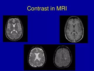
International Journal of Trend in Scientific Research and Development (IJTSRD) @ www.ijtsrd.com eISSN: 2456-6470 techniques in this study, based on two parameters of PSNR and MSE. The primary aim of enhancing contrast is to bring out detailed information obscured in an image. For the analysis purpose, various contrast enhancement techniques are used including Contrast Stretching, Shock Filter, Histogram Equalization, Contrast Limited Adaptive Histogram Equalization CLAHE. These techniques are applied on the three types of brain MRI images, which are normal, benign and malignant brain MRI images [2]. Based on two evaluation parameters Peak signal to noise ratio (PSNR) and Mean square error (MSE) the comparison was made between the techniques [4], [11]. It also has been described which technique is best suited for brain MRI analysis and gives better performance than others give. A.Contrast Stretching Contrast stretching is a straightforward method for enhancing the image contrast by extending the scope of pixel intensity values to expand the parameter set necessary. This methodology can only apply a linear scaling function to the pixel values for images [6]. Through contrast stretching a low-contrast image may be converted into a high-contrast image by restoring or remapping the gray-level values to the full range of the histogram. [13]. It is referred to as dynamic range extension in the scope of digital signal processing. This can be demonstrated in equation (1): < ≤ + − L x b , y b x b γ Where, x and y are input image and the Stretched output respectively and α, β and γ represents stretching constants, acting as factor of multiplier whereas the lower and the higher range are represented by a and b while are calculated by equation (2) and (3): with a minimum principle which keeps the range of the filtered image surrounded by the original image range [8]. The dilation and erosion process is expounded pursuant to a diminutive time increment dt using a Partial Differential Equation (PDE), this creates a sharp discontinuity called a borderline shock around two areas of influence and ultimately we get a deblurred output. The Kramer and Bruckner definition [7] can be describe utilizing this subsequent Partial Differential equation (4): ( ) gradient . u delta sign ut= Considering a continuous image ( under the process may generate a class of filtered images. The equation (4) can be written as equation (5): ( u sign ut ∆ ∆ − = Where subscript denotes partial derivatives, and ( y u u = ∇ is the gradient ofu . Let's assume some pixels are in the maximum influence zone (negative Laplacian i.e. yy xx u u u + = ∆ , is negative which will then an equation (6) is given for the dilation. u ut ∇ = For Laplacian positives, pixels pertain to a minimum zone of influence, with 0 < ∆u , then (5) can be abridged to an erosion given by equation (7). u ut ∇ − = Therefore the Laplacian's zero-crossings dole out as an edge detector. Essentially, the enhancement/sharpening of image input. C.Histogram Equalization Typically a histogram represents uniform distribution of pixels in a graphical form. The histogram equalization (HE) is a widely used image contrast enhancement technique because of its simplicity and efficacy [9, 13]. This method improves the global image contrast and accommodates image intensities to boost contrast by distributing the intensity levels that are most widely used [4], thus the intensities of the histogram are better distributed. Thus, the areas with lower local contrast reach a greater contrast [3], [10]. Considering a discrete gray scale image comprising of L discrete gray levels expressed as{ 1 2 1 0 − L , , X ...... X X X . Over a certain image X , the probability density function equation (8): ( n Where, the numbering of occurrence of gray level Xis represented by Defining the cumulative distribution function (cdf) consequent to ( K X P is mentioned as the following equation (9): ( ) ( ) u (4) ) y f x, . Then, evolving f ) u (5) )T xu , (6) (7) α ≤ < 0 , x β x a ( ( ) = − + (1) y x a y , a x b a ) consequence is an ≤ < y and b y a = α ya a (2) ( ) = β − + y b a y (3) b a The intent of stretching the contrast in the different applications is to introduce the image into a scope which is more acquainted or normal to the senses, it is therefore also called normalization [5]. B.Shock Filter For deblurring signals and images shock filter is used by creating shocks at points of inflection. Shock filters either apply erosion or dilation process produces a "shock" between two zones of influence, one is for a maximum and the other for a minimum signal. The premise is that the step of dilation is utilised near a maximum, and the process of erosion is used in the vicinity of minimum. The determination about pixel's area of persuade (whether maximum or minimum) is constructed on the Laplacian basis as the pixel is perceived to be a maximum for negative Laplacian in the zone of influence, and minimum for positive Laplacian. Shock filters comply { ( j , i ) } X = X } ( ) is determined as in P X K n ) k k= P X (8) kn , n is total pixel count in the image. ) @ IJTSRD | Unique Paper ID – IJTSRD30336 | Volume – 4 | Issue – 3 | March-April 2020 Page 177
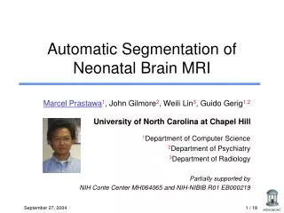
International Journal of Trend in Scientific Research and Development (IJTSRD) @ www.ijtsrd.com eISSN: 2456-6470 − − 1 1 k m n ( ) 1 ∑ = j ∑∑ = 0 i ) j , i is noise free [ ] ( j , i ) ( j , i ) ( ) x 2 = = − C P X MSE x y j mn = 0 0 j 1 , and (9) . (12) gray scale image and ( . ) j , i Where ( is noisy approximation of ( 3. Correlation Coefficient -The correlation coefficient is a function of correlation connecting two variables, which ranges from –1 to 1. The correlation coefficient will be either 1 or –1 where the two variables are in perfect linear connection. The sign varies depending on how the variables are related either positively or negatively. If there is no linear relation between variables, the correlation coefficient is 0. Here two dissimilar types of coefficients for correlation are taken into account; first one is the Pearson product- moment correlation coefficient which is more widely used in measuring the association between two variables, and the other being the Spearman rank correlation coefficient, which is based on the rank relationship between variables. Given paired measurements (X1, Y1), (X2, Y2), (Xn, Yn), the Pearson product moment measurement is specified by equation (13): ( ) 1 ≤ = − m× ) j , i x ≤ 0 1 Where x is Histogram equalization is a mapping mechanism mapping the input image throughout the dynamic range ( using the cdf as a level transformation function. A transformation function ( ) x f based on the cdf is defined as in equation (10): ( ) C . X X X x f L 0 1 0 − + = − This is the required Histogram Equalization output image. D.Contrast Limited Adaptive Histogram Equalization (CLAHE) Contrast Limited Adaptive Histogram Equalization (CLAHE) is an Adaptive Histogram Equalization generalization which is used to avert noise amplification problems [6]. This CLAHE algorithm separates the images into contextual regions and contributes to the equalization of the histograms to each [4]. It evinces the distribution of used gray values and makes the features of hidden images more evident. The approach comes with three parameters: ?Block size: This represents the scale of the local region that equalizes the histogram around a pixel. This scale should be greater than the preservable characteristics. ?Histogram bins: Histogram bins that are utilized in numbers for the process of histogram equalization. The number of pixels in a block should be smaller than that. This value also limits output quantification when processing RGB images of 8 bit gray, or 24 bit. ?Max slope: It restricts the stretch of contrast in the feature to pass strength. High local contrast can result in very large values. The process takes one added ' clip-level ' parameter, which varies from 0 to 1. This methodology calculates the histogram for each pixel, and after that performs the equalization operation of the window or block size. III. EVALUATION OF PERFORMANCE MATRICES The two metrics are utilized to evaluate the performance of different methods of enhancement are considered below [4, 11]. 1. PSNR - The peak signal-to-noise ratio, abbreviated as PSNR, is the ratio of the maximum signal power to the power of corrupting noise affecting the fidelity of its representation. PSNR can be represented by equation (11). Here, I Max is the maximum possible pixel value of the image. The specimens are shown using linear PCM with B bits per sample, I Max is 1 2 − 2. MSE -The Mean Square error abbreviated as MSE provides the cumulative squared error between the original image and its noisy approximation. The lower the value of MSE, the lower the error. MSE is given below by equation (12): X for k ,.... L x y n 0 C X k k ) by X ,X − 0 1 L ( ) ( ) x (10) correlation coefficient (13) IV. Dataset and Software Implementation The data set used for the testing and information on the implementation of the program are given in the following subsections. Experiments were performed to evaluate different commonly used enhancement techniques for different type of diseased brain MRI images by the comparison we can find the best suited method for enhancement of Brain MRI image. Dataset In the current study, 81 patients dataset constituting of 11 Benign, 25 Gliomas, 30 Meningioma and 15 Metastases, are taken from 512 MR brain tumor slices marked by the radiologists using CBAC out of which four images from each category is shown in the fig. 1 to fig. 4 respectively. These images are collected online available dataset from the website radiopedia.org. RESULTS AND ANALYSIS 2 Max I = 10 PSNR . log 10 MSE (11) (a) B . (b) @ IJTSRD | Unique Paper ID – IJTSRD30336 | Volume – 4 | Issue – 3 | March-April 2020 Page 178
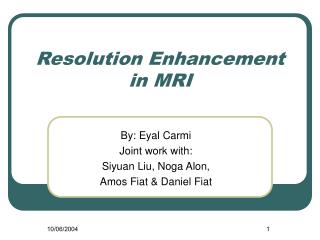
International Journal of Trend in Scientific Research and Development (IJTSRD) @ www.ijtsrd.com eISSN: 2456-6470 (d) (c) Fig. 2 (a), (b), (c) & (d) are different types of Gliomas Brain MRI images (d) Fig.1 (a), (b), (c) & (d) are different types of Benign Brain MRI images (a) (b) (a) (c) (b) (d) (c) Fig. 3 (a), (b), (c) & (d) are different types of Meningioma Brain MRI images @ IJTSRD | Unique Paper ID – IJTSRD30336 | Volume – 4 | Issue – 3 | March-April 2020 Page 179
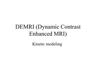
International Journal of Trend in Scientific Research and Development (IJTSRD) @ www.ijtsrd.com eISSN: 2456-6470 (c) (a) (d) Fig. 4 (a), (b), (c) & (d) are different types of Metastases Brain MRI images (b) Software Implementation These proposed methods are implemented in MATLAB 9.0 and are tested on various brain tumor MR images of size 400×400. The experiments were performed on PC having Intel™ i3 Processor 3.0 GHz processor with 4 GB RAM. The algorithm takes 3 min for training the samples. The comparison of various contrast enhancement techniques for gray scale images is carried out based on the two parameters that are Peak Signal to Noise Ratio (PSNR) and Mean Square Error (MSE). These parameters are being used as objective measures for evaluating the performance of the improvement methods applied. As per the evaluation, the result of normal, benign and malignant brain MRI images are mentioned in following Table I, Table II and Table III. TABLEI OUTCOME OF DISTINCT ENHANCEMENT TECHNIQUES FOR BENIGN BRAIN MRI IMAGE Parameters PSNR Image Category Technique Applied MSE 2051.840 15.0358 0.51651 9253.218 8.94204 0.58559 CC Contrast Stretching Shock Filter Histogram Equalization 234.0376 26.8647 0.21827 CLAHE 995.266 TABLEIII OUTCOME OF DISTINCT ENHANCEMENT TECHNIQUES FOR GLIOMAS BRAIN MRI IMAGE Benign Brain MRI Image 18.3353 0.81775 Parameters PSNR Image Category Technique Applied MSE CC Contrast Stretching Shock Filter Histogram Equalization 425.00703 23.6879 0.21827 CLAHE 1287.6206 17.2889 0.82576 1047.2492 18.1140 0.41745 4928.6040 11.4430 0.34158 Gliomas Brain MRI Image TABLEIIIII OUTCOME OF DISTINCT ENHANCEMENT TECHNIQUES FOR MENINGIOMA BRAIN MRI IMAGE Parameters PSNR Image Category Technique Applied MSE 862.0564 19.0362 0.74360 5672.74 11.6601 0.27683 CC Contrast Stretching Shock Filter Histogram Equalization 1073.816 18.7656 0.35151 CLAHE 1293.88 TABLEIVV OUTCOME OF DISTINCT ENHANCEMENT TECHNIQUES FOR METASTASES BRAIN MRI IMAGE Meningioma Brain MRI Image 17.6669 0.85294 Parameters PSNR Image Category Technique Applied MSE CC Contrast Stretching Shock Filter Histogram Equalization 881.30001 20.4975 0.28053 CLAHE 3178.0222 17.6209 0.88397 1364.4298 16.9439 0.29657 5011.9875 11.3849 0.52930 Metastases Brain MRI Image @ IJTSRD | Unique Paper ID – IJTSRD30336 | Volume – 4 | Issue – 3 | March-April 2020 Page 180
International Journal of Trend in Scientific Research and Development (IJTSRD) @ www.ijtsrd.com eISSN: 2456-6470 [6]R. C. Gonzalez and R. E. Woods, Digital Image Processing, 2nd ed. Englewood Cliffs, NJ: Prentice-Hall; 2002 V. In this research paper, multiple contrast enhancement techniques are applied for brain MRI image analysis and comparison was made, which is useful in determining the best method for clinical diagnosis. It is clear from the above comparison tables that the Histogram Equalization filter gives the minimum MSE and the highest PSNR value and best Correlation coefficient; therefore, it is the best suited method and has delivered better performance than others. Consequently, this provides sonologist and radiologist better visual perception for the diagnostic purpose of brain MRI disease. We will be applying other Biomedical Image Processing approaches for better performance in future. References [1]Masalkar D N, Shitole A S. Advance Method for Brain Tumor Classification. International Journal on Recent and Innovation Trends Communication 2014; 2(5): 1255-9. CONCLUSION [7]A. K. Jain, Fundamentals of Digital Image Processing., Englewood Cliffs, NJ: Prentice Hall; 1989. [8]V. Shrimali, R. S. Anand and V. Kumar, “Comparing the Performance of Ultrasonic Liver Image Enhancement Techniques: A Preference Study,” IETE Journal of Research, Vol. 56, Issue 1, Jan-Feb 2010. [9]W. M. Hafizah and E. Supriyanto, “Comparative Evaluation of Ultrasound Kidney Image Enhancement Techniques,” International Journal of Computer Applications (0975 – 8887), Vol. 21, No. 7, May 2011. [10]S. S. Al-amri, N. V. Kalyankar and S. D. Khamitkar., “Linear and Non-linear Contrast Enhancement Image,” International Journal of Computer Science and Network Security (IJCSNS), Vol. 10, No. 2, February 2010. in Computing and [2]Sharma Y, Chhabra M. An Improved Automatic Brain Tumor Detection System. International Journal of Advanced Research in Computer Science and Software Engineering 2015; 5(4): 11-5. [11]J. Weickert, “Coherence-Enhancing Shock Filters,” Pattern Recognition, Springer- 2003. [12]C. Ludusan, O. Lavialle, S. Pop, R. Terebes and M. Borda, “Image Enhancement Using a New Shock Filter Formalism”, Acta Technica Napocensis, Electronics and Telecommunications, Vol. 50, No. 3, 2009. [3]Mansa S M, Kulkarni N J, Randive S N. Review on Brain Tumor Detection and Segmentation Techniques. International Journal of Computer Applications 2014; 95: 34-8 [13]Y. T. Kim, “Contrast Enhancement Using Brightness Preserving Bi-Histogram Transactions on Consumer Electronics, Vol. 43, No. 1, February 1997. [4]Anandgaonkar G, Sable G. Brain Tumor Detection and Identification from T1 Post Contrast MR Images Using Cluster Based Segmentation, InternationalJournal of Science and Research 2014; 3(4): 814-7. Equalization,” IEEE [14]H. Yeganeh1, A. Ziaei and A. Rezaie, “A Novel Approach for Contrast Enhancement Based on Histogram Equalization,” Proceedings of the International Conference on Computer and Communication Engineering 2008, Kuala Lumpur, Malaysia, 978-1- 4244-1692-9/08 ©2008 IEEE. [5]Sadi, A.; Elmoataz, A.; Toutain, M. Nonlocal PDE morphology: A generalized shock operator on graph. Signal Image Video Process. 2016, 10, 439–446. @ IJTSRD | Unique Paper ID – IJTSRD30336 | Volume – 4 | Issue – 3 | March-April 2020 Page 181