The Brain in MRI and CT
430 likes | 729 Views
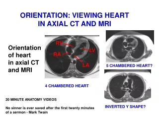
The Brain in MRI and CT. MRI are taken by a rotating magnetic field CT scans are taken by rotating X-ray source. Always Your Right is the Patient’s Left. Coronal. Axial. Patient. You. Patient.

The Brain in MRI and CT
E N D
Presentation Transcript
The Brain in MRI and CT • MRI are taken by a rotating magnetic field • CT scans are taken by rotating X-ray source
Always Your Right is the Patient’s Left Coronal Axial Patient You Patient In axial MRI , you looking from down to top, as if you look to the patient from the feet “see demonstration” In coronal MRI, you looking to the patient face to face. You
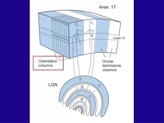
Ventral “What” pathway • Carries information about static object properties such as colour, luminance, stereopsis and pattern recognition. • Slow pathway from P-ganglion cells (through laminae 3-6 of LGN, V1) to V2, V4 and inferior temporal cortex
Dorsal “Where” pathway • Information about dynamic object properties- motion and spatial relationships • Fast pathway for transient visual signals • Pathway to V1, V2, MT, medial superior temporal and parietal lobe
Damage to “What” pathway Achromatopsia, agnosia
Achromatopsia • Complete achromatopsia- BL area V4: Lingual/fusiform gyri/occipitotemporal junction
Color agnosia • Color agnosia: loss the ability to retrieve color knowledge • cannot name colors for objects but can sort • Remembering the color of object • Color composition Left or bilateral occipitotemporal region Inferior temporal , fusiform and right lingual
Color anomia • Inability to name colors or to point to colors given their names, which is not due to aphasia or due to defective color perception
Color anomia • Inability to name colors or to point to colors given their names, which is not due to aphasia or due to defective color perception • Usually associated with left mesial occipitotemporal region • hence usually affect the visual cortex or optic rediation leading to right hemianopia , and also associated with alexia
The Neural Basis of Visual Perception • Visual agnosia is the inability to recognize objects despite satisfactory vision. • Caused by damage to the pattern pathway usually in the temporal cortex. • For words : Alexia
Agnosia • Topographagnosia • Inability to navigate routes using familiar landmarks - deficit in familiar scene perception • Right lingual gyrus • Alexia • Left (dominant lobe) fusiform/lingual areas
Lesion, left occipitotemporal region and involves parts of the lingual and fusiform gyri. Hemi- achromatopsia , pure alexia , and category-specific visual object agnosia
Occipitotemporal gyri
Occipitotemporal gyri
Prosopagnosia- Inability to recognize or learn faces Identify people by other cues- gait, mannerisms or facial features- spectacles, gait Aware of defect BL lingual and fusiform gyri of medial occipitotemporal cortex. Agnosia