Download
1 / 5
100 likes | 633 Views
Brain MRI in Multiple Sclerosis. Anne G. Osborn, M.D. Professor of Radiology University of Utah School of Medicine. Figure 2 MRI Axial FLAIR scan showing white matter foci of increased signal intensity surrounding cavitating areas characteristic of long-standing MS .

E N D
Brain MRI in Multiple Sclerosis Anne G. Osborn, M.D. Professor of Radiology University of Utah School of Medicine
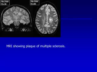
Figure 2 MRI Axial FLAIR scan showing white matter foci of increased signal intensity surrounding cavitating areas characteristic of long-standing MS.
Figure 3 MRI axial FLAIR scan of deep periventricular foci of increased signal intensity surrounding cavitating areas.
Figure 4 MRI sagittal FLAIR scan with classic calloseptal deep periventricular foci perpendicular to ventricle surface classic for Dawson fingers.