Learning objectives :
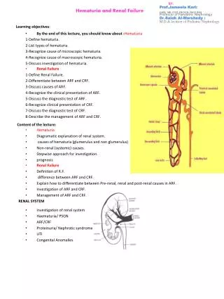
BY: Prof. Jameela Kari: CABP, MD, CCST, FRCPCH, FRCP (UK) Professor of Paediatric Nephrology Dr. Salah Al- Morshedy : M.D.& lecturer of Pediatric Nephrology. Hematuria and Renal Failure. Learning objectives :. Renal Failure 1-Define Renal Failure.

Learning objectives :
E N D
Presentation Transcript
BY:Prof.Jameela Kari:CABP, MD, CCST, FRCPCH,FRCP (UK)Professor of Paediatric NephrologyDr.Salah Al-Morshedy :M.D.& lecturer of Pediatric Nephrology Hematuria and Renal Failure Learning objectives: • Renal Failure 1-Define Renal Failure. 2-Differentiate between ARF and CRF. 3-Discuss causes of ARF. 4-Recognize the clinical presentation of ARF. 5-Discuss the diagnostic test of ARF. 6-Recognize clinical presentation of CRF. 7-Discuss the diagnostic test of CRF. 8-Describe the management of ARF and CRF. • By the end of this lecture, you should know about :Hematuria 1-Define hematuria. 2-List types of hematuria. 3-Recognize cause of microscopic hematuria. 4-Recognize cause of macroscopic hematuria. 5-Discuss investigation of hematuria. Content of the lecture: • Hematuria • Diagramatic explanation of renal system. • causes of hematuria (glumerulus and non glumerulus) • Non-renal (systemic) causes. • Stepwise approach for investigation. • prognosis • Renal Failure • Definition of R.F. • difference between ARF and CRF. • Explain how to differentiate between Pre-renal, renal and post-renal causes in ARF. • Investigation of ARF and CRF. • Management of ARF and CRF. RENAL SYSTEM • Investigation of renal system • Haematuria/ PSGN • ARF/CRF • Proteinuria/ Nephrotic syndrome • UTI • Congenital Anomalies
Investigation of the renal system HAEMATURIA • Urine analysis • Urine electrolytes: fraction excretion of sodium (pre-renal <1%, renal >1-2%) • 24 hours urine for protein, Ca etc • GFR (creatinine clearance= ml/1.73m2/minute) • Cultures: older children: midstream Younger children:SPA, Catheter, Bag • Imaging: US, KUB/IVU, MCUG, CT scan, isotope scan (DMSA, DTPA) • Renal biopsy • Macroscopic = frank = gross • Microscopic > 5 RBCs per high powered field • Red urine: • Blood (RBCs= haematuria, haemoglobin= haemoglobinuria) • Myoglobin • Food: blackberries • Aniline dyes used for coloring candy • Drugs: Rifampicin, phenolphthalein • Urate • Porphyrins Causes • Glomerular: brownish or cola-coloured and may contain RBCs cast, proteinuria • Lower urinary tract: red to pink color urine and may contain clots PATHOPHYSIOLOGY • Haematological: thrombocytopaenia, DIC, coagulopathies, renal vein thrombosis, SCD • Glomerular: brownish or cola-coloured and may contain RBCs cast, proteinuria • Stones and hypercalciuria • Infection: bacterial, TB, viral • Lower urinary tract: red to pink color urine and may contain clots • Anatomic abnormalities: congenital, trauma, polycystic kidneys, tumors, vascular abnormalities • Exercise and drugs Glomerular Diseases • 7 years old boy • Frank haematuria (smokey or tea colored) • H/O throat infection 2 weeks ago • O/E peri-orbital edema, BP 140/90 (hypertension) • What is the most likely diagnosis • What investigations Post-streptococcal glomerulonephritis: • Rare before the age of 3 years • Nephritic picture: Gross haematuria, edema , hypertension, renal insufficiency (normal RF-ARF) • Complications of hypertension: encephalopathy, congestive heart failure • Rarely: nephritic- nephrotic picture.
Diagnosis: Complications: ARF volume overload, hypertension, fits, hyperkalemia, hyperphosphatemia, hypocalcaemia and acidosis • Antibiotics • Management of ARF Treatment • Urine analysis: RBCs, RBCs cast, proteinuria • Low complement C3 • Evidence of streptococcal infection: throat culture, ASO titer and DNase B antigen and streptozyme test • Mild normochromic anaemia • Renal function Prognosis • Complete recovery 95% • Infrequently: severe acute phase leading to chronic renal insufficiency • Recurrence are extremely rare Glomerular Diseases • MPGN • Rapidly progressive glomerulonephritits • SLE • Shunt nephritis • Goodpasture disease • Membranous • Anaphlactoidpurpura • IgA nephropathy • Idiopathic (benign familial) • Alport syndrome • RBCs cast • Glomerulonephritis or vasculitis • Exclude extra-renal disorders EVALUATION OF A CHILD WITH HAEMATURIA • History • Examination • Investigation: • Studies performed in all patients: • urine microscopy and culture • CBC • serum creatinine • serum C3 level • US kidneys • urine protein = urine albumin/creatinine ratio • calcium = urine calcium/creatinine ratio
Studies performed in selected patients: • Dnase B titer or streptozyme < 6 months duration • Skin or throat culture • ANA titer • Urine analysis looking for cast • Coagulation study/ platelet count • Sickle cell screen in black patients • Audiogram • Renal biopsy • Microscopic haematuria plus any of the following: • Diminished renal function • Proteinuria • Persistant microscopic haematuria (>1 year) • Second episode of gross haematuria • Cystoscopy • Pink o microscopic haematuria, dysuria and sterile urine culture Case History: • 5 years old boy • Generalized malaise, abdominal pain, joints pain, peri orbital oedoma Henoch-Schönlein Purpura or (Anaphylactoid Purpura) • Renal involvement occurs in 25–50% of children during the acute phase • Haematuria with or without casts or proteinuria during the first few weeks of illness • The nephrotic syndrome, moderate azotemia, hypertension, oliguria, and hypertensive encephalopathy may occasionally occur. • Most children with renal involvement recover Recurrent Gross Haematuria or Persistent Microscopic Haematuria • IgA nephropathy (Berger) • Alport syndrome • Familial idiopathic haematuria • Idiopathic hypercalciuria. IgA Nephropathy (Berger) • Glomerulonephritis with IgA as the predominant immunoglobulin in mesangial deposits, in the absence of any systemic disease • Haematuria + minimal proteinuria • Normal C3 + usually normal RF • Diagnosis: renal biopsy IgA Nephropathy (Berger) • Treatment: supportive • Prognosis: mainly good, only 30% has progressive disease:hypertension, diminished renal function, or proteinuria exceeding 1 g/24 hr between episodes of gross hematuria
ALPORT Syndrome. Idiopathic Familial Benign Haematuria • No proteinuria • All investigations normal • Urine test of the parents and siblings • An excellent prognosis, but long-term follow-up is required to exclude Alport syndrome Idiopathic Hypercalciuria • Hereditary nephritis. • Haematuria + proeinuria + sensorineural hearing loss (minority) + eye abnormalitie (10%). • Diagnosis: renal biopsy. • Males with Alport syndrome commonly develop end-stage renal failure in the 2nd or 3rd decade of life, occasionally in association with hearing loss. Females usually have a normal life span and only subclinical hearing loss. • RGH, persistent microscopic hematuria, or dysuria in the absence of stone formation • Hypercalciuria (without hypercalcemia) • Diagnosis: 24-hr urinary calcium excretion exceeding 4 mg/kg, urine calcium to creatinine ratio (mg/mg) • Hypercalciuria may lead to nephrolithiasis • RX: Oral thiazide Membranous Glomerulopathy • Uncommon in childhood and a rare cause of haematuria. • The most common cause of nephrotic syndrome in adults. • Associated with systemic lupus erythematosus, cancer, gold or penicillamine therapy, and syphilis and hepatitis B virus infections. MEMBRANOPROLIFERATIVE (MESANGIOCAPILLARY) GLOMERULONEPHRITIS • Chronic glomerulonephritis that frequently leads to glomerular destruction and end-stage renal failure. • Most common in the second decade of life. • Presentation: nephrotic syndrome, gross hematuria or asymptomatic microscopic hematuria, proteinuria and hypertension . renal function may be normal to depressed. Low C3 complement level. • Diagnosis by renal biopsy. GLOMERULONEPHRITIS OF CHRONIC INFECTION • Subacute bacterial endocarditis (S. viridans and other organisms) • Infected ventriculoatrial shunts for hydrocephalus (Staphylococcus epidermidis) • Syphilis, hepatitis B, hepatitis C, candidiasis, and malaria • Immune complexes, which deposit in the kidneys and initiate the glomerulonephritis. • The C3 level is frequently depressed. RAPIDLY PROGRESSIVE (CRESCENTIC) GLOMERULONEPHRITIS • Nephritis with rapid progression to end-stage renal failure • Causes: poststreptococcal, lupus, membranoproliferative, and the glomerulonephritides of Goodpasture disease, anaphylactoidpurpura, and other forms of vasculitis • Acute renal failure, often after an acute nephritic or nephrotic episode • Diagnosis: Renal biopsy • Paediatric Nephrology Emergency Acute Renal Failure • Develops when renal function is diminished to the point at which body fluid homeostasis can no longer be maintained. • Oliguria (daily urine volume less than 400 ml/m2) is common, the urine volume may approximate normal. • Nonoliguric renal failure: in certain types of acute renal failure (aminoglycosidenephrotoxicity).
Etiology • Prerenal causes • Hypovolemia, hypotension, hypoxia • Renal causes • Acute tubular necrosis • Acute interstitial nephritis • Glomerulonephritis • Localized intravascular coagulation • Tumors • Developmental abnormalities • Hereditary nephritis • Postrenal causes • Obstructive uropathy, stone, blood clot CLINICAL MANIFESTATIONS • Diminished urine output • Oedema (salt and water overload) • Hypertension, vomiting, and lethargy (uremic encephalopathy). • Complications of acute renal failure: volume overload with congestive heart failure and pulmonary edema, arrhythmias, gastrointestinal bleeding due to stress ulcers or gastritis, seizures, coma, and behavioral change • Life threatening: GIT bleed, pericarditis and encephalopathy Diagnosis • Careful history • Examination • Investigation: CBC, urea and electrolytes, PO4, Ca, blood gases, C3, US kidneys, urine electrolytes ( Na and creatinine), fractional excretion of Na (less than 1% in hypovalaemia) Urine Analysis Prerenal. • Urine osmolality exceeds 500mOsm/kg [mmol/l] H2O. • Sodium content is usually less than 20mEq/l (mmol/l). • The fractional excretion of sodium (urine/plasma sodium concentration divided by the urine/plasma creatinine concentration X 100) is usually less than 1%. Renal • osmolality less than 350 mOsm/kg [mmol/l] H2O • Usually exceeds 40 mEq/l (mmol/l) • Usually exceeds 1% Treatment • Pre-renal=Hypovolemia: volume replacement may be critical • Renal: • Fluid restriction: input= output + 400 ml/m2/24 hr (insensible losses) • Hyperkalemia: no potassium-containing fluid, foods, or medications until adequate renal function is re-established • > 7 mEq/L (mmol/L): Nebulisedsalbutamul, IV Calcium gluconate, sodium bicarbonate, ca resonium, glucose and insulin • Moderate acidosis is common in renal failure: Na bicarbonate Treatment • Hypocalcemia and hyperphosphataemia: Ca binders (Ca carbonate). • Hypertension: • The primary disease process (nifedipine, diazoxide, sodium nitroprusside or labetalol as a continuous intravenous infusion is indicated for hypertensive crises). • Expansion of the extracellular fluid volume (salt and water restriction is critical). • Indications for dialysis: fluid overload, and congestive heart failure, electrolyte abnormalities (especially hyperkalemia), central nervous system disturbances, hypertension.
Prognosis HEMOLYTIC-UREMIC SYNDROME • Frequently follows an episode of gastroenteritis caused by an enteropathogenic strain of Escherichia coli (0157:H7). It has been associated with other bacterial (Shigella, Salmonella, Campylobacter, S. pneumoniae), Bartonella, and viral • Most common in children under the age of 4 yr • usually preceded by gastroenteritis (fever, vomiting, abdominal pain, and diarrhea, which is often bloody) or, less commonly, by an upper respiratory tract infection. • In general, recovery of function is likely following renal failure resulting from prerenal causes, the hemolytic-uremic syndrome, acute tubular necrosis, acute interstitial nephritis, or uric acid nephropathy. • On the other hand, recovery of renal function is unusual when renal failure results from most types of rapidly progressive glomerulonephritis, bilateral renal vein thrombosis, or bilateral cortical necrosis. Hemolytic-Uremic Syndrome • Followed in 5–10 days by the sudden onset of pallor, irritability, weakness, lethargy, and oliguria. • Physical examination may reveal dehydration, edema, petechiae, hepatosplenomegaly, and marked irritability. • Diagnosis: microangiopathic hemolytic anemia, thrombocytopenia, and acute renal failure. Chronic Renal Failure • In children under 5 yr of age is commonly the result of anatomic abnormalities (hypoplasia, dysplasia, obstruction, malformations) • After 5 yr of age acquired glomerular diseases (glomerulonephritis, hemolytic-uremic syndrome) or hereditary disorders (Alport syndrome, cystic disease) predominate • UT malformation + Glomerulonephritis + Pyelonephritis….. > 50% of causes MANAGAMENTdepends upon the degree of renal insufficiency (CRD) STAGES OF C.R. DISEASE
Clinical Manifestations Management • Nonspecific symptoms (headache, fatigue, lethargy, anorexia, vomiting, polydipsia, polyuria, growth failure). • Physical examination: pallor and weakness, hypertension, growth retardation and rickets. • Investigations: • CBC: anaemia. • Electrolytes: hyponatremia, hyperkalemia, acidosis. • BUN and creatinine (nitrogen accumulation and level of renal function). • hypocalcemia, hyperphosphatemia, osteodystrophy. • High of intact parathyroid hormone levels. • Diet in chronic renal failure. • Water and electrolyte management in chronic renal failure (fluid, K, Na). • Acidosis in chronic renal failure. • Renal Osteodystrophy. • Anemia in chronic renal failure. • Hypertension in chronic renal failure. • Drug dosage in chronic renal failure. End-stage Renal Failure • Dialysis is generally initiated when the patient's GFR < 10 ml/1.73m2/minute • Continuous ambulatory peritoneal dialysis (CAPD) • Continuous cyclic peritoneal dialysis (CCPD) • Haemodialysis • Haemofiltration • Renal transplantation APD=CCPD Peritoneal Dialysis=CAPD
Haemodialysis Peritoneal Dialysis Haemodialysis