Download

1 / 20
200 likes | 687 Views
Hypersensitivity. Ali Al Khader, M.D. Faculty of Medicine Al-Balqa’ Applied University Email: ali.alkhader@bau.edu.jo. Introduction. = immunologically mediated tissue injury 4 types: I II III

E N D
Hypersensitivity Ali Al Khader, M.D. Faculty of Medicine Al-Balqa’ Applied University Email: ali.alkhader@bau.edu.jo
Introduction = immunologically mediated tissue injury • 4 types: I II III IV These are the topics that we will discuss today
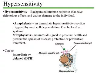
Immediate (Type I) hypersensitivity May include glandular secretions Systemic or local
Immediate (Type I) hypersensitivity • = allergy • Routes of re-exposure: -Injection…like bee sting -Ingestion…like peanut allergen -Inhalation -Direct contact
Immediate (Type I) hypersensitivity…late phase • Stays for days without additional antigen exposure • Especially in asthma and allergic rhinitis • Infiltration of tissues with eosinophils, neutrophils, basophils, monocytes, and CD4+ T cells • Tissue destruction, typically in the form of mucosal epithelial cell damage
Some molecules and cells in type I hypersensitivity • IL-13…IgE production …mucus secretion • Mast cells are abundant around vessels and nerves and subepithelial • Some mast cell secretagogues: -IL-8 -codeine -morphine -adenosine -melittin…in bee venom -physical stimuli • Mast cell will secrete: -preformed (primary) mediators -secondary mediators (synthesized de novo) • Histamine also increases mucus production in nasal, bronchial and gastric mucosae
Some molecules and cells in type I hypersensitivity, cont’d • Mast cell enzymes: -tissue damage -generation of kinins -activate complement precursors • Mast cell proteoglycans…store amines • Prostaglandin D2 also increases mucus secretion • PAF…a lipid mediator but not from arachidonic acid …vasodilation, permeability and bronchospasm …platelet aggregation and release of histamine
Non-atopic immediate (Type I) hypersensitivity • 20-30% of type I hypersensitivity reactions are not triggered by antigens …but induced by temperature changes or exercise without Th2 or IgE contribution …mainly an abnormality in mast cell response … = non-atopic allergy
Antibody-mediated (Type II) hypersensitivity mechanisms • IgG on cells opsonization • IgG & IgM on cells classical pathway C3b & C4b opsonization • IgG & IgM on cells classical pathway membrane attack complex • Antibody-dependent cellular cytotoxicity (ADCC): …IgG on cells binding to Fc by phagocytes NK cells
Antibody-mediated (Type II) hypersensitivity mechanisms, cont’d • Antibodies deposited on fixed tissues complement activation C3a & C5a (anaphylatoxins) …also WBCs binding through their Fc & C3b receptors enzymes & ROS chemotaxis of neutrophils & monocytes -Some forms of glomerulonephritis -Vascular rejection in organ grafts Inflammation
Antibody-mediated (Type II) hypersensitivity mechanisms, cont’d • Cellular dysfunction without injury or inflammation …Myasthenia gravis …Graves disease
Examples of type II hypersensitivity • Transfusion reactions…preformed antibodies • Hemolytic disease of the newborn (erythroblastosis fetalis) …IgG antierythrocyte antibodies from the mother cross the placenta and cause destruction of fetal red cells • Autoimmune hemolytic anemia, agranulocytosis and thrombocytopenia • Certain drug reactions…act as haptens that bind to plasma proteins on RBCs
Immune complex-mediated (Type III) hypersensitivity • Systemic immune complex disease mechanisms: -Acute serum sickness is the prototypical example …large amounts of foreign serum (e.g., serum from immunized horses used for protection against diphtheria) -Often glomeruli and joints are affected
Immune complex-mediated (Type III) hypersensitivity • Local immune complex disease mechanisms: = Arthus reaction -acute & local -usually in the skin -mainly experimentally induced -injection of antigen (sensitization) dermal injection of that antigen the antigen gains access to local blood vessels complexes are formed causing vasculitis with fibrinoid necrosis of that vessel followed by ischemia and tissue necrosis locally
T cell–mediated (Type IV) hypersensitivity • CD4+ T cells…cytokines or • CD8+ T cells…killing -induced by environmental and self antigens -the cause of many chronic inflammatory diseases, including autoimmune diseases Delayed-type hypersensitivity reaction is the prototype Th1 & Th17 -DM type 1 -important in graft rejection
CD4+ T Cell–Mediated Inflammation, sensitization • Dendritic cell + antigen + CD4+ T cell IL-2 more CD4 activation • Cytokines from APCs differentiation to Th1 and Th17 by IFN-gamma & IL-12 by IL-1, IL-6 and IL-23 Now we have memory pool of T cells
CD4+ T Cell–Mediated Inflammation, repeated exposure • IFN-gamma by Th1 classical activation of macrophages • In addition to the actions of these macrophages, they augment Th1 responses by IL-12 and by expressing more MHC II • Th17 secrete: -IL-17 -IL-22 -chemokines -IL-21…amplifies Th17 responses recruitment of neutrophils and monocytes
CD4+ T Cell–Mediated Inflammation, repeated exposure • Tuberculin test is the prototypical example • Granuloma and perivascular cuffing by mononuclear cells (CD4+ cells and macrophages) are typical histological patterns here • Contact dermatitis is also an example Skin rashes in most drug reactions Similar mechanisms