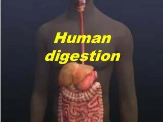
HUMAN DIGESTION
HUMAN DIGESTION. Digestion. Digestion is the breakdown of large, complex organic molecules into smaller components that can be used by the body. Molecules need to be small enough to diffuse across plasma membranes. Four Components of Digestion.

HUMAN DIGESTION
E N D
Presentation Transcript
Digestion • Digestion is the breakdown of large, complex organic molecules into smaller components that can be used by the body. • Molecules need to be small enough to diffuse across plasma membranes.
Four Components of Digestion • Ingestion – this is the consumption of or taking in of nutrients. • Digestion – the chemical breakdown of large organic molecules into smaller components by enzymes. • Absorption – the transport or delivery of digested nutrients to body tissues. • Egestion – the elimination of food waste materials from the body.
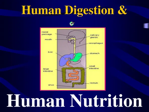
Ingestion • Food enters the human digestive tract through the mouth or oral cavity. • Humans are considered chunk feeders because they consume chunks of food that are then mechanically broken down.
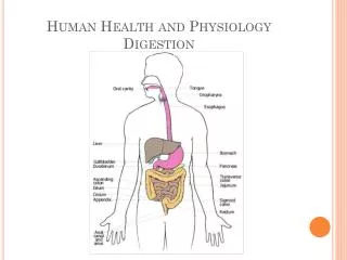
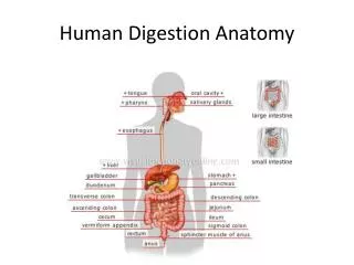
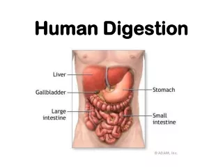
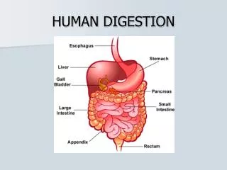
Alimentary Canal • The human digestive tract is often referred to as the alimentary canal. • The alimentary canal of a normal adult is approximately 6.5 to 9 meters long.
Mechanical Digestion • Physical breakdown of food begins with the teeth grinding the food and increasing its surface area. An increase surface area allows for easier chemical digestion • Bacteria living in the mouth can feed off of nutrients sticking to the teeth and cause tooth decay.
Saliva • Saliva is released from the salivary glands and begins chemical digestion of starches. Saliva contains the enzyme salivary amylase which breaks down starches into simpler carbohydrates.
Taste Buds • As the food particles dissolve in the saliva they penetrate the cells of the taste buds located on the tongue and cheeks. • Humans can differentiate between sweet, sour, salty and bitter.
Swallowing • Saliva also lubricates the food and helps to form a bolus, the ball of food that is swallowed.
Esophagus • The bolus of food moves down the esophagus propelled by wave-like muscular contractions known as peristalsis. • Peristalsis moves food all the way through the gastrointestinal tract.
Stomach • The stomach acts as a temporary storage site for food. Food usually spends about 4 hours in the stomach. It has ridges which allow it to expand to store about 1.5 litres of food. • The stomach is also the site of initial protein digestion.
Stomach • Movement of food into and out of the stomach is controlled by circular muscles known as sphincters. • One at the top of the stomach allows food from the esophagus to enter and prevents food from going back up into the esophagus. • Another located at the bottom slowly releases partially digested food into the small intestine. The partially digested food is called chyme.
Stomach • Millions of cells lining the stomach secrete various fluids known collectively as gastric fluids. • Gastric fluid consists of mucus, hydrochloric acid, pepsinogens and other substances. • Mucus coats and protects the lining of the stomach. Hydrochloric acid kills any harmful substances that have been ingested and it also converts pepsinogen into pepsin. • Pepsin is a protein digesting enzyme that breaks large protein chains into smaller chains.
Stomach pH • The pH environment of the stomach normally ranges between 2.0 and 3.0 on the pH scale. • The high acidity allows pepsin to work and makes the HCL effective at killing pathogens
Stomach Ulcers • An stomach ulcer is a lesion in the lining of the stomach. It occurs when the protective mucus lining breaks down and the cell membranes are exposed to the HCl and pepsin • Most stomach ulcers are linked to the bacterium shown on the right known as Heliobacter pylori.
Endoscopy • An endoscope (shown on the right) can be used to view things such as stomach ulcers or as shown below, a tumor growing in the large intestine. • The endoscope can also extract small pieces of tissue for a biopsy.
Small and Large Intestine • The intestines are named for their diameter, not length. • The small intestine is up to 7 m in length but only 2.5 cm in diameter. • The large intestine is only 1.5 m in length but 7.6 cm in diameter.
Small Intestine • In mammals the length of the small intestine is directly related to their diet. • Due to the fact that meats are easier to digest than plant materials, carnivores (lion) will have a shorter intestine than herbivores (rabbit). The length of the digestive tract of omnivores falls somewhere in the middle.
Anatomy of the Small Intestine • The majority of chemical digestion occurs in the first of three sections of the small intestine known as the duodenum. • This section also contains an opening from the bile duct and pancreatic duct through which bile and pancreatic enzymes enter the small intestine.
Small Intestine • Food enters the small intestine as a semi-solid mixture known as chyme. The chyme is acidic due to the HCl in the stomach so it needs to be neutralized. • The presence of chyme in the small intestine triggers the conversion of prosecretin into secretin which is absorbed into the blood stream and carried to the pancreas
Pancreas • The pancreas is an accessory organ of the digestive system. It releases chemicals to aid in digestion. • Secretin will stimulate the pancreas to release a solution containing bicarbonate ion into the small intestine where it will neutralize the acidic chyme and raise the pH from 2.5 to 9.0. This inactivates the pepsin.
Pancreas and Digestion • The pancreas also releases digestive enzymes that break down the three macromolecules: carbohydrates, lipids and proteins.
Pancreas & Proteins • Trypsinogen, a protein-digesting enzyme is released into the small intestine where it is convertes into trypsin and it breaks down large protein chains into smaller chains. • The final step in protein digestion occurs with the release of erepsins from the pancreas and they break the smaller chains into individual amino acids.
Pancreas and Carbohydrates • Amylase enzymes are released from the pancreas that break large carbohydrate chains into small chains called disaccharides. • Then the small intestine releases disaccharide enzymes which break those small chains into individual sugars.
Pancreas and Lipids • The pancreas also releases enzymes known as lipases that break down fats into fatty acids and glycerol. • The lipases include pancreatic lipase and phospholipase. • Before lipids can be broken down by lipases they must first be emulsified.
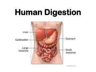
Liver • The liver is a large accessory organ of the digestive system that is constantly producing a fluid known as bile. • Bile is stored in the gall bladder until it is needed in the small intestine.
Liver and Gall Bladder • The presence of lipids in the small intestine trigger the release of the hormone cholecystokinin (CCK) which triggers the release of bile from the gall bladder. • Bile contains bile salts that emulsifies fats which means it breaks them into smaller droplets so they can be digested.
Gallstones • Bile contains cholesterol which can acts as a binding agent and cause bile salts to crystallize into gallstones. • Gallstones can block the bile duct and inhibit fat digestion while causing a lot of pain.
Jaundice • Jaundice is a yellow discoloration of the skin and tissues caused by a collection of bile pigments in the blood. • The pigments are a result of the liver breaking down hemoglobin from red blood cells and the products are stored in the gall bladder.
Detoxification and the Liver • The liver is also able to detoxify many substances in the body by making them soluble and they can then be dissolved in the bloodstream and eliminated in urine. • One example would be alcohol. Alcohol can damage liver cells which are replaced by connective tissues and fat. The result is cirrhosis of the liver (shown left).
Absorption of Materials • Chemical digestion of nutrients is completed by the time it reaches the large intestine. • Now that nutrients are small enough they need to be absorbed into the blood stream so they can diffuse inside cells.
Large Intestine • The large intestine or colon stores waste products long enough so that water can be reabsorbed from the wastes. • Along with the water, some inorganic salts, minerals and vitamins are absorbed.
Large Intestine • The large intestine is home to several different types of bacteria. • These bacteria use waste materials to synthesize vitamins B and K. This is an example of a symbiotic relationship.
Cellulose • Cellulose is a long chain carbohydrate found in the cell wall of plant cells. • Humans cannot digest cellulose however it provides bulk which promotes the movement of the waste products out of the colon.
Cellulose • Cellulose is more commonly known as fiber. Fiber helps to remove wastes and therefore toxins from the body. • If you have a diet low in fiber you will have fewer bowel movements which means toxins remain in your body for longer periods of time.
Absorption of Nutrients • Most nutrients are absorbed in the small intestine. • The small intestine is lined with millions of small finger-like projections known as villi. The villi increase the surface are of the small intestine which increases it’s ability to absorbed digested nutrients.
Structure of the Villus • Each villus contains a capillary network along with a lacteal. • End products of protein and carbohydrate digestion enter the capillary network. • End products of fat digestion are absorbed into the lacteal. The lacteal is a vessel of the lymphatic system.