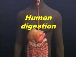
Human Physiology: Digestion
Human Physiology: Digestion. Topic 6.1, 11.3 Option H2, H3, H4. 3 Dietary Categories. Herbivores Cattle, gorillas, snails, and sea urchins, eat autotrophs (plants and algae) Carnivores Lions, hawks, spiders, and snakes Ingest other animals Omnivores

Human Physiology: Digestion
E N D
Presentation Transcript
Human Physiology:Digestion Topic 6.1, 11.3Option H2, H3, H4
3 Dietary Categories • Herbivores • Cattle, gorillas, snails, and sea urchins, • eat autotrophs (plants and algae) • Carnivores • Lions, hawks, spiders, and snakes • Ingest other animals • Omnivores • Crows, cockroaches, raccoons, and humans • Ingest both plants and animals
How do animals obtain and ingest their food? • Suspension feeders • Extract food particles suspended in the surrounding water • Ex. Clams and oysters • Substrate feeders • Live on or in their food source and eat their way through it. • Ex. Caterpillars and earthworms • Fluid feeders • Obtain food by sucking nutrient nutrient-rich fluids from a living host, either a plant or an animal. • Ex. Mosquitoes and ticks • Bulk feeders • Ingest relatively large pieces of food • Ex. most animals
Overview: Food processing • Four stages • Ingestion The act of eating • Digestion The breaking down of food into molecules small enough for the body to absorb. Two phases: 1. Breaking food down mechanically (teethchewing) into smaller pieces 2. hydrolysis, chemical breakdown, catalyzed by enzymes • Absorption Cells lining the digestive tract take up (absorb) the products of digestion—small molecules such as amino acids and simple sugars Nutrients travel through blood to cells, where they are made into macromolecules or further broken down for energy • Elimination undigested material passes out of the digestive tract
General compartments for digestion • Food vacuoles are the simplest digestive compartments. • Phagocytosis: cell engulfs food particle, which then fuses with a lysosome. Most animals have an alimentary canal, a digestive tube with two openings, a mouth and an anus. • Allows food to move in one direction, with specialized regions in the digestive tube that carry out digestion and absorption of nutrients in sequence
General compartments for digestion • Food entering the mouth usually passes into: • A pharynx, or throat • Then passes into the esophagus • Passed to stomach, muscular and churns and grinds food • Chemical digestion and nutrient absorption occur mainly in the intestine • Undigested materials are expelled though the anus.
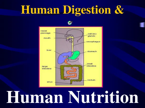
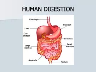
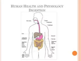
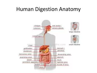
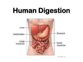
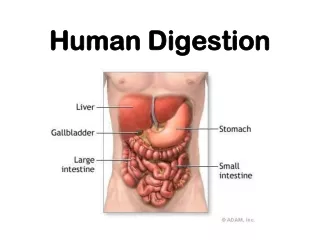
Human Digestive Tract • Main parts of human alimentary canal: • Mouth, oral cavity, tongue, pharynx, esophagus, stomach, small intestine, large intestine, rectum, and anus. • Main digestive glands • Salivary glands, pancreas, and liver • Secrete digestive juices that enter the alimentary canal through ducts. • Secretions from liver are stored in gallbladder before they are released into the intestine.
Human Digestive Tract • Peristalsis • Rhythmic waves of contractions of smooth muscles in the walls of the digestive tract • Once food is swallowed, peristalsis propels it through the alimentary canal. • In only 5-10 seconds, food passes from the pharynx down the esophagus and into the stomach. • Pyloric sphincter, a muscular ringlike valve, keeps food in the stomach by regulating the pass of food into the small intestive. • Works like a drawstring, closing off the tube and keeping food in the stomach long enough for stomach acids and enzymes to begin digestion. • http://nutrition.jbpub.com/resources/animations.cfm?id=1&debug=0
Human Digestive Tract • Final steps of digestion and nutrient absorption occur in the small intestine over a period of 5-6 hours. • Undigested material moves slowly through the large intestine (taking 12-24 hours), and feces are expelled through the anus.
Digestion Begins in the Oral Cavity • Saliva is produced by the salivary glands through ducts to the oral cavity even before you eat; it’s a response to presence of food • Sight or smell of food causes nerve stimulation • In a typical day, salivary glands secrete more than a liter of saliva
Digestion Begins in the Oral Cavity • Saliva contains several substances necessary for food processing • Slippery glycoproteins • Protects the soft lining of the mouth and lubricates food for easier swallowing • Buffers • Neutralize food acids, helping prevent tooth decay. • Antibacterial agents • Kill many of the bacteria that enter the mouth with food. • Salivary amylase • Digestive enzyme that begins hydrolyzing food
Digestion Begins in the Oral Cavity • Oral Cavity • Mechanical and chemical digestion begins in the oral cavity. • Chewing cuts, smashes, and grinds food, making it easier to swallow and exposing more food surface to digestive enzymes • Teeth and tongue are prominent • Teeth grind and crush food • Tongue, muscular organ with taste buds, allows you to taste your meal and manipulates food and helps shape it into a ball called a bolus. • In swallowing, it pushed the bolus to the back of the oral cavity and into the pharynx.
Pharynx- Swallowing • Pharynx has openings for both the esophagus and the trachea (wind-pipe) • Most of the time, esophageal opening is closed and air enters the trachea and proceeds to the lungs. • When you eat: • a bolus of food enters the pharynx, triggering the swallowing reflex • The esophageal sphincter relaxes and allows the bolus to enter the esophagus • Larynx (voice box) moves upwards and tips the epliglottis over the tracheal opening. • Epiglottis prevents food from passing into the trachea. • After the bolus enters the esophagus, the larynx moves downward, the epiglottis moves up again, and breathing passage reopens • Esphogas sphincter contracts above the bolus.
Esophagus: Peristalsis • Esophagus is a muscular tube that conveys food boluses from the pharynx to the stomach. • Muscles at the very top of esophagus are under voluntary control; thus, the act of swallowing begins voluntarily. • Then, Involuntary waves of contraction by smooth muscles in the rest of the esophagus take over.
Esophagus: Peristalsis • As food is swallowed, muscles above the bolus contract, pushing the bolus downward • Simultaneously, muscles around the bolus relax, allowing the passageway to open. • Muscle contractions continue in waves until the bolus enters the stomach. • Waves of smooth muscle contraction also move materials through small and large intestine
Stomach: stores and breaks down food • Stomachs are the main reason we do not have to eat constantly • Highly elastic and can stretch to accommodate about 2 Liters of food and drink, usually enough to satisfy our body’s needs for many hours. • Some digestion occurs in the stomach. • The stomach secrete gastric juice: • made up of mucus, enzymes, and strong acid. • Functions to break apart the cells in food • Also kills most bacteria and other microbes that are swallowed with food.
Stomach: stores and breaks down food • Stomach wall is highly folded, and has pits that lead to tubular gastric glands. • Three types of cells that secrete different components of the gastric juice: • Mucous cells: secrete mucous, which lubricates and protects the cells lining the stomach • Parietal cells: secrete HCl acid • Chief cells: secrete pepsinogen, an inactive form of the enzyme pepsin
Stomach: stores and breaks down food • Pepsinogen, HCl, and pepsin: • 1. Pepsinogen and HCl are secreted into the lumen (cavity) of the stomach. • 2. HCl converts pepsinogen to pepsin. • 3.Pepsin then activates more pepsinogen, starting a chain reaction. Pepsin begins the chemical digestion of proteins. Proteins will be further digested in small intestine.
Stomach: stores and breaks down food • Prevention of gastric juice from digesting away stomach lining: • Secreting pepsin in the inactive form of pepsinogen helps protect the cells of the gastric glands • mucus helps protect the stomach lining from both pepsin and acid. • Still, epithelium is constantly eroded; enough new cells are generated by mitosis to replace the stomach lining completely about every three days.
Stomach: stores and breaks down food • Gastric glands are regulated by a combination of nerve signals and hormones: • When you see, smell, or taste food, a signal from your brain to your stomach stimulates your gastric glands to secrete gastric juice. • Once food is in your stomach, substances in the food stimulate cells in the stomach wall to release the hormone gastrin in the circulatory system. • Gastrin circulates in the blood stream, returning to the stomach wall, stimulating further secretion of gastric juice. • As much as 3L of gastric juice may be secreted a day. • A negative-feedback mechanism inhibits secretion of gastric juice when the stomach contents become too acidic. • Acid inhibits the release of gastrin, and with less gastrin in the blood, the gastric glands secrete less gastric juice.
Stomach: stores and breaks down food • About every 20 seconds, the stomach contents are mixed by the churning action of muscle in the stomach wall and result in acid chyme. • Opening between the esophagus and the stomach is usually closed until a bolus arrives. • Backflow of acid chyme causes heartburn (which should be called esophagus-burn) • Can also cause acid-reflux (gastroesophageal reflux disease; GERD)
Stomach: stores and breaks down food • Pyloric sphincter helps regulate the passage of acid chyme from the stomach into the small intestine. • The stomach takes about 2-6 hours to empty after a meal; acid chyme leaves stomach only a squirt at a time. • Acid chyme rich in fats stimulates the small intestine to release a hormone that slows the emptying of the stomach, providing more time for digestion. • Other hormones secreted by the small intestine influence the release of digestive juices from the pancreas and gall bladder.
Gastric Ulcers • Gastric Ulcers: • Open sores that form when mucus, which normally protects the stomach wall from the corrosive effect of digestive juice, fails to protect it. • Small intestine and esophagus are also susceptible to ulcers • Symptoms are usually gnawing pain in the upper abdomen, which may occur a few hours after eating. • Were formerly thought to result from the production of too much pepsin/and or acid or too little mucus: • For years, the blame was put on factors that cause these effects, such as aspirin, ibuprofen, smoking, alcohol, coffee, and stress • However, strong evidence now points to…
Gastric Ulcers • H. pylori • A spiral-shaped bacteria • Low pH of the stomach kills most microbes, but not this one! • Burrows beneath mucus and releases harmful chemicals • Growth seems to result in a localized loss of protective mucus and damage to the cells lining the stomach • WBC fight infection, causing mild inflammation of the stomach, called gastritis. • Gastric ulcers form when pepsin and HCl destroy cells faster than the cell can regenerate from the H. pylori attack. • Eventually, stomach will erode to the pint where it actually has a hole in it, which can lead to a life threatening infection in abdomen or internal bleeding. • 70-90% of ulcer and gastritis sufferers have this bacterial infection • Also found in 30% of healthy people. linked to certain kinds of stomach cancer
Gastric Ulcers • Treatment • Usually respond to a combination of anti-biotics and bismuth (the active ingredients of Pepto-Bismol) which eliminates bacteria and promotes healing. • Drugs that reduce stomach acidity may also help, and researchers are working on preventitive treatment for H.Pylori.
Small Intestine • Once at the S.I., food has been mechanically reduced to smaller pieces and mixed with juices; it now resembles a thick soup. • Starch digestion began in the mouth, and protein breakdown began in the stomach. • All other chemical digestion occurs in the s.i. • Nutrients are also absorbed into the blood from the s.i. • Length of over 6m, making it the longest organ of the alimentary canal. Diameter is only about 2cm, which is why it’s called the “small” intestine.
Small Intestine • Contributing to digestion in s.i. are two large glandular organs: pancreas and liver. • Pancreas: • Produces pancreatic juice • a mixture of digestive enzymes and an alkaline solution rich in bicarbonate • Alkaline solution neutralizes acid chyme as it enters the small intestine
Small Intestine • Liver • Performs a wide variety of functions, including the production of bile: • Contains bile salts that emulsify fats, making them more susceptible to attack by digestive enzymes. • Gall bladder stores bile until it is needed in the small intestine.
Small Intestine • First 25 cm or so of the s.i. is called the duodenum. • Where acid chyme squirted from the stomach mixes with bile from the gall bladder, pancreatic juice from the pancreas, and digestive enzymes from gland cells in the in the intestinal wall.
Small Intestine • All four types of macromolecules (carbohydrates, proteins, nucleic acids, and fats) are digested. • Refer to table 21.11 on pg. 438
Small Intestine • Carbohydrate digestion: • Begins in the oral cavity and is completed in the s.i. • Pancreatic amylase hydrolyzes starch (a polysaccharide) into the disaccharide maltose • Maltose is then hydrolyzed into glucose via maltase. • Sucrase hydrolyzes table sugar and lactase digests milk sugar (lactose)
Small Intestine • Protein digestion: • S.i. completes protein digestion from the stomach • Pancreas and duodenum secrete hydrolytic enzymes that completely dismantle polypeptides into amino acids. • dipeptidase • Hydrolyzes fragments only two or three amino acids long. • trypsin and chymotripsin • Break polypeptides into smaller polypeptides • Trypsinogen (in pancreas) is converted into trypsin by the action of enteropeptidase (the enzyme that is bound to the membranes of the small intestine). • aminopeptidase and carboxypeptidase • Split off one amino acid at a time, working from both ends of a polypeptide.
Small Intestine • Nucleic acid digestion: • Nucleases hydrolyzes the nucleic acids in food. • From the pancreas • Split DNA and RNA (which are present in the cells of food items) into their component nucleotides, which are then broken down into nitrogenous bases, sugars, and phosphates by other enzymes produced by the duodenal cells.
Small Intestine • Fat digestion: • Most fat remains undigested until it reaches the duodenum. • Hydrolysis of fats is problematic due to fats insolubility in water. Emulsification= Problem Solved!!! • Bile salts in bile cause fat globules to be physically broken up into smaller fat droplets, a process called emulsification. • Many small droplets allows for a larger surface area of fat exposed to lipase, an enzyme that breaks fat molecules down into fatty acids and glycerol.
Small Intestine • Problems with lipid digestion in a hydrophillic medium: • Lipids tend to coalesce (lump together) and are only accessible to lipase at the lipid-water interface. • Bile molecules have a hydrophobic end and a hydrophilic end which emulsifies (prevents from coalescing) the lipids • Lipase must be water-soluble and has a hydrophobic active site (for its substrate, lipids) • The increased surface area allows lipase greater access to its substrate
Small Intestine • By the time persistalsis has moved the mixture of chyme and digestive juices through the duodenum, chemical digestion of your meal is just about complete. • Main function of the rest of the small intestine is the absorption of nutrients and water.
Small Intestine • Structurally, great for nutrient absorption. • Lining has a huge surface area– roughly 300 m2, about the size of a tennis court • Extensive surface area results from several kinds of folds and projections. • Villi: large circular folds with numerous, small fingerlike projections around the inner wall of the s.i. • Microvilli: many tiny surface projections found on epithelial cells lining a villus. • extend into the lumen of the intestine and greatly increase the surface area across which nutrients are absorbed.
Small Intestine • Some nutrients are absorbed via simple diffusion; other nutrients are pumped against concentration gradients into the epithelial cells • The core of each villus is penetrated by a small lymph vessel and a network of capillaries. • After fatty acids and glycerol are absorbed by an epithelial cell, these building blocks are recombined into fats and are transported into the lymph vessel. • Amino acids and sugars pass out of the intestinal epithelium and then across the thin walls of the capillaries into the blood.
Small Intestine • Capillaries that drain nutrients away from the villi converge into larger veins and eventually into a main vessel, the hepatic portal vein, that leads directly to the liver: • Liver gets first access to nutrients absorbed from a meal • Converts many nutrients into new substances that the body needs. • One of its main functions is to remove excess glucose from the blood and convert it to glycogen, which is stored in liver cells. • From the liver, blood travels to the heart, which pumps the blood and the nutrients it contains to all parts of the body.