The Lymphatic System
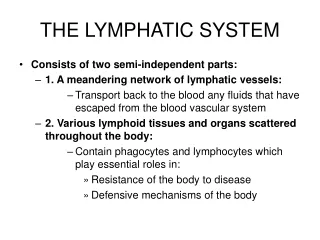
The Lymphatic System. Chapter 21. Introduction. The lymphatic system supports the function of the cardiovascular and immune systems of the body The lymphatic system consists of two semi-independent parts A network of lymphatic vessels Lymphoid organs scattered throughout the body


The Lymphatic System
E N D
Presentation Transcript
The Lymphatic System Chapter 21
Introduction • The lymphatic system supports the function of the cardiovascular and immune systems of the body • The lymphatic system consists of two semi-independent parts • A network of lymphatic vessels • Lymphoid organs scattered throughout the body • The lymphatic vessels transport fluids that have escaped from the cardio-vascular system
Lymphatic Vessels • As blood circulates through the body, exchanges of nutrients, wastes, and gases occur between the blood and the interstitial fluid • The fluid that remains behind in the tissue spaces, as much as 3 liters a day, become part of interstitial fluid
Lymphatic Vessels • These leaked fluids, as well as any plasma proteins that escape from the blood-stream, must be carried back to the blood if the cardiovascular system is to sufficient blood volume to operate properly • The lymphatics are elaborate system of drainage vessels that collects the excess protein-containing interstitial fluid and returns it to the bloodstream • Once interstitial fluid enters the lymphatics ducts it is called lymph
Distribution of Lymphatic Vessels • The lymphatic vessels form a one-way system in which lymph flows only toward the heart • The system begins with the lymph capillaries
Distribution of Lymphatic Vessels • Lymph capillaries weave between the tissue cells and blood capillaries in the loose connective tissue of the body
Distribution of Lymphatic Vessels • Lymph capillaries are widespread, occurring almost everywhere blood capillaries occur • Lymph capillaries are absent from bone and teeth, bone marrow, and the entire central nervous system
Distribution of Lymphatic Vessels • Although similar to blood capillaries, lymphatic capillaries are remarkably permeable • The great permeability is due to structural modifications • Minivalves • Anchoring filaments
Minivalves • The endothelial cells forming the walls of the lymph capillaries are not tightly joined; instead their edges loosely overlap forming easily opened, flaplike minivalves
Anchoring Filaments • Bundles of fine filaments anchor the endothelial cells to surrounding structures so that any increase in interstitial fluid volume separates the cell flaps, exposing gaps in the wall and allowing fluid to enter rather than the capillary collapsing
Lymphatic Vessels • These structural modifications create a system where the valves gap open when fluid pressure is greater in the interstitial space, allowing fluid to enter the lymphatic capillary • Pressure inside the lymphatic capillary forces the minivalve flaps together prevent-ing a leak back out
Lymphatic Vessels • Proteins present in the interstitial fluid are prevented from entering the blood capillaries but enter lymphatic capillaries • In addition, when tissues are inflamed, lymphatic capillaries develop openings that permit uptake of even larger particles such as cell, pathogens, bacteria, viruses, and cancer cells • Thus cancer cells can use lymphatic capillaries to travel throughout the body
Lymphatic Vessels • Highly specialized lymphatic capillaries called lacteals are present in the fingerlike villa of the intestinal mucosa • The lymph draining from the digestive viscera is milky white rather than clear because the lacteals also receive digested fat from the intestine • This creamy lymph, called chyme, is also delivered to the blood via the lymphatic system • This concept discussed further in Chap 24
The Lymphatic System • From the lymphatic capillaries, lymph flows through successively larger channels • Collecting vessels • Trunks • Ducts
The Lymphatic System • Collecting vessels have the same three tunics as veins, but they are thinner-walled, have more internal valves, and anastomose more • In general the collect- ing vessels in the skin travel along with superficial veins of the CV system while deep vessels of the trunk travel with arteries
The Lymphatic System • The lymphatic trunks are formed by the union of the largest collecting vessels, and drain fairly large areas of the body • The trunks are named for the areas from which they collect lymph • Lumbar • Bronchomediastinal • Subclavian
The Lymphatic System • Lymph is delivered to one of two large ducts in the thoracic region • The right lymphatic duct drains lymph from the upper arm and the right side of the head and thorax • The larger thoracic duct receives lymph from the rest of the body
The Lymphatic System • Each terminal duct empties the lymph into the venous circulation at the junction of the internal jugular vein on its side of the body
Lymph Transport • Unlike the cardiovascular circulation, the lymphatic system lacks an organ that acts as a pump • Under normal conditions, lymphatic vessels are very low pressure conduits • Compression of skeletal muscle, pressure changes associated with respiration and valves to prevent back flow, aid the movement of lymph • Smooth muscle in the lymphatic duct contracts rhythmically to move lymph along
Lymph Transport • About 3 liters of lymph enters the blood- stream every 24 hours, a volume that almost equal to the amount of fluid lost to the tissue spaces from the bloodstream in the same time period • Movement of the adjacent tissues are extremely important in propelling lymph through the lymphatics • Physical activity or passive movement increase lymph flow
Lymphoid Cells • In order to understand some of the basic aspects of the lymphatic system’s role in body protection and immunity it is necessary to understand the components • Lymphoid cells • Lymphoid tissues
Lymphoid Cells • Infectious microorganisms, such as bacteria and viruses, that manage to penetrate the body’s epithelial barrier begin to quickly proliferate in the underlying loose tissue • These invaders are fought off by the inflammatory response by phagocytes (macrophages) and lymphocytes
Lymphoid Cells • Lymphocytes, the main warriors of the immune system, arise in red bone marrow • They then mature into one of the two main varieties of immunocompetent cells • T cells (T lymphocytes) • B cells (B lymphocytes) • These cells act to protect the body against antigens (bacteria and their toxins, viruses, mismatched RBC’s, or cancer cells
Lymphoid Cells • Activated T cells manage the immune response and some of them directly attack and destroy foreign cells • B cells protect the body by producing plasma cells, daughter cells that secrete antibodies into the blood • Antibodies immobilize antigens until they can be destroyed by phagocytes
Lymphoid Cells • Lymphoid marcophages play a crucial role in body protection and in the immune response by phagocytizing foreign substances and helping to activate T cells • Dendritic cells found in lymphoid tissue also activate T cells • Reticular cells are fibroblast cells that produce the reticular fiber stroma or network that supports the other cells types in the lymphoid organs
Lymphoid Tissue • Lymphoid tissue is an important component of the immune system because it • Houses and provides a proliferation site for lymphocytes • Furnishes an ideal surveillance vantage point for both lymphocytes and macrophages
Lymphoid Tissue • Lymphoid tissue, a type of loose connective tissue called reticular connective tissue, dominates all lymphoid organs except the thymus • The dark staining areas represent the connective tissue fibers
Lymphoid Tissue • Macrophages live on the fibers of the network • Within the spaces of this network are huge numbers of lymphocytes Macrophage Lymphocytes Reticular fiber
Lymphoid Tissue • Lymphocytes squeeze through the walls of capillaries and venules to reside temporarily in the lymphoid tissue and then leave to patrol the body • The cycling of lymphocytes between the circulatory vessels, lymphoid tissues, and loose connective tissues of the body ensures that lymphocytes reach infected or damaged sites quickly
Lymphoid Organs • Lymphoid organs as exemplified by lymph nodes, the spleen, and the thymus are discrete collections of lymphoid tissue • The exact pattern of the lymphoid tissue differs in the various lymphoid organs
Lymphoid Organs • Lymphoid organs are discrete, encapsulated collections of diffuse lymphoid tissue and nodules • The exact pattern of lymphoid tissue differs in the various lymphoid organs
Lymph Nodes • As lymph is transported back to the bloodstream, it is filtered through lymph nodes that cluster along the lymphatic vessels of the body
Lymph Nodes • There are hundreds of lymph nodes that are usually imbedded in connective tissue an not seen • Large clusters of lymph nodes occur near the body surface in the inguinal, axillary, and cervical regions of the body • Located where vessels form large trunks
Lymph Nodes • Lymph nodes have two basic functions, both concerned with body protection • They act to filter lymph • Phagocytic macrophages in the nodes remove and destroy microorganisms and other debris that enter the lymph from the loose connective tissue, effectively preventing further spread • They play a role in activating the immune system • Lymphocytes in the lymph nodes monitor the lymphatic stream for the presence of antigens and attack them
Lymph Nodes • Lymph nodes are small (2.5 cm), bean shaped structures surrounded by a fibrous capsule of connective tissue
Lymph Nodes • Trabecula are connective tissue strands that extend inward to divide the node into compartments
Lymph Nodes • Its internal of framework of reticular fibers physically supports the ever-changing population of lymphocytes
Lymph Nodes • Two histologically distinct regions in a lymph node are the cortex and the medulla • These areas contain densely packed follicles with dividing B cells Cortex Medulla
Lymph Nodes • The outer cortex contains densely packed follicles, many with germinal centers heavy with dividing B cells
Lymph Nodes • Dendritic cells nearly encapsulate the follicles and abut the rest of the cortex, which primarily houses T cells in transit • The T cells circulate continuously between the blood, lymph nodes, and lymphatic stream, performing their surveillance role
Lymph Nodes Medullary cords • Medullary cords are thin inward extensions of the cortex containing lymphocytes and plasma cells
Lymph Nodes • Throughout the node are lymph sinuses which are large lymph capillaries spanned by reticular fibers • Numerous marcophages reside on these reticular fibers and phagocytize foreign matter in the lymph as it flows by the sinuses • Lymph borne antigens in the lymph leak into the surrounding reticular tissue, where they activate some of the strategically positioned lymphocytes to mount an immune response
Circulation in Lymph Nodes • Lymph enters the convex side of a lymph node through a number of afferent lymphatic vessels
Circulation in Lymph Nodes Subcapsular sinus • Lymph moves through a large, baglike sinus, the sub- capsular sinus, into a number of smaller sinuses that cut through the cortex and enter the medulla
Circulation in Lymph Nodes • Lymph meanders through these sinuses and finally exits the node at its hilus, via efferent lymphatic vessels
Circulation in Lymph Nodes • Because there are fewer efferent vessels draining the node than there afferent vessels feeding it, the flow of lymph through the node stagnates somewhat, allowing time for the lymphocytes and macrophages to carry out their protective functions • In general, lymph passes through several nodes before its cleansing process is completed
Lymph Nodes: Clinical • Inflammation of a node is caused by a large number of bacteria trapped in a node • Inflammation results in swelling and pain • Lymph nodes can become secondary cancer sites, particularly in metastasizing cancers that enter lymphatic vessels and become trapped • Cancer infiltrated nodes are swollen but not painful
Other Lymphoid Organs • Lymph nodes are just one type of many types of lymphatic tissue • Other lymphoid organs include • Spleen • Thymus gland • Tonsils • Peyer’s patches
Other Lymphoid Organs • The common feature of all lymphoid organs is that they are all composed of reticular connective tissue • Additionally, all lymphoid tissues help protect the body
Spleen • The soft, blood rich spleen is about the size of fist and is the largest lymphoid organ