Parasitology Sporozoons
1.26k likes | 1.37k Views
Learn about the classification, life cycles, and clinical importance of sporozoons. Explore different flagellates like Giardia and Trichomonas, with symptoms, diagnosis, and treatments. Understand infections like Leishmania and Trypanosoma.

Parasitology Sporozoons
E N D
Presentation Transcript
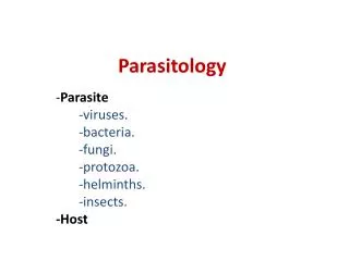
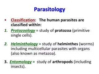
Parasitology/Sporozoons (3 hours) • 1. Defines “Sporozoon’’ • 1.1 Lists the sporozoon classification. • 1.2. Defines the cell structure of sporozoons. • 1.3. Defines the life cyles of sporozoons; lists the cysts, trophozoits, oocysts, promastigot, amastigot structures. • 2. Lists the clinical tables related with sporozoons and defines pathogenetic mechanisms. • 2.1. Defines the clinical importance of sporozoons. • 2.2. Defines the sample taking related with infections of sporozoons. • 2.3. Lists the laboratory diagnostic methods.
Intestinal flagellates Giardia intestinalis Trichomonas spp Dientamoeba fragilis Trichomonas vaginalis Hemoflagellates Leishmania donovani Trypanosoma cruzi Flagellates
Giardia intestinalis (lamblia) • Cysts are resistant forms and are responsible for transmission of giardiasis. • Both cysts and trophozoites can be found in the feces (diagnostic stages) • The cysts are hardy and can survive several months in cold water. • Infection occurs by the ingestion of cysts in contaminated water, food, or by the fecal-oral route (hands or fomites) • Because the cysts are infectious when passed in the stool or shortly afterward, person-to-person transmission is possible
Symptoms • diarrhea, • abdominal pain, • bloating, • nausea, • vomiting. incubation period: 1 to 14 days (average of 7 days)
Diagnosis • cysts or trophozoites in the feces, using direct mounts (repeated samplings) • samples of duodenal fluid (e.g., Enterotest) • duodenal biopsy may demonstrate trophozoites • alternate methods • antigen detection tests enzyme immunoassays, • detection of parasites by immunofluorescence.
Trichomonas vaginalis • resides in the female lower genital tract and the male urethra and prostate • transmitted among humans, its only known host, primarily by sexual intercourse • no cyst formation
Symptoms • vaginitis with a purulent discharge is the prominent symptom, • vulvar and cervical lesions, • abdominal pain, • dysuria • dyspareunia. In men • asymptomatic; • urethritis, • epididymitis, • prostatitis the incubation period is 5 to 28 days.
Diagnosis • microscopic examination of wet mounts • culture
Dientomoeba fragilis • not an ameba but a flagellate • no cyst formation
Symptoms • diarrhea, • abdominal pain, • anorexia, • nausea, • vomiting, • fatigue, • weight loss.
Diagnosis • detection of trophozoites in permanently stained fecal smears (e.g., trichrome). • not detectable by stool concentration methods. • trophozoites can be easily overlooked ,they are pale-staining and their nuclei may resemble those of Endolimax nana or Entamoeba hartmanni.
Leishmania donovani • a vector-borne disease • transmitted by sandflies • caused by obligate intracellular protozoa of the genus Leishmania
Promastigotes • amastigotes • in the sandfly's midgut, the parasites differentiate into promastigotes in Mexico Central America, South America southern Europe Asia (not Southeast Asia), the Middle East, and Africa
Symptoms Cutaneous leishmaniasis (L.tropica) • one or more sores on their skin. • the sores can change in size and appearance over time. • they often end up looking somewhat like a volcano, with a raised edge and central crater. • a scab covers some sores. • the sores can be painless or painful. • some people have swollen glands near the sores (for example, in the armpit if the sores are on the arm or hand).
Symptoms Visceral leishmaniasis (kala-azar)(L.donovani) • fever, • weight loss, • enlarged spleen and liver (usually the spleen is bigger than the liver). • Some patients have swollen glands. • Certain blood tests are abnormal. low blood counts, including a low red blood cell count (anemia), low white blood cell count, and low platelet count. • Some patients develop post kala-azar dermal leishmaniasis. • Visceral leishmaniasis is becoming an important opportunistic infection in areas where it coexists with HIV.
Diagnosis • Giemsa-stained slides • culture (using for example the diphasic NNN medium) • Antibody detection
Trypanosoma cruzi • Chagas disease, • a zoonotic disease • transmitted to humans • by blood-sucking triatomine bugs (kissing bug) • Chronic Chagas disease is a major health problem in many Latin American countries
trypomastigotes • amastigotes • trypomastigotes • amastigotes Trypanosoma cruzi can also be transmitted through blood transfusions, organ transplantation, transplacentally, in laboratory accidents.
Symptoms • fever, • anorexia, • lymphadenopathy, • mild hepatosplenomegaly, • myocarditis. Chronic stage may not occur for years or even decades after initial infection. • cardiomyopathy (the most serious manifestation); • megaesophagus • megacolon; • weight loss. Chronic Chagas disease and its complications can be fatal.
Diagnosis Microscopic examination: • of fresh anticoagulated blood, or its buffy coat, for motile parasites; • of thin and thick blood smears stained with Giemsa, for visualization of parasites. Isolation of the agent by: • inoculation into mice; • culture in specialized media (e.g. NNN, LIT); • xenodiagnosis, where uninfected reduviid bugs are fed on the patient's blood, and their gut contents examined for parasites 4 weeks later.
Treatment • benznidazole or nifurtimox • In the chronic stage, e.g., pacemaker for heart block
Trypanosoma brucei • T. b. gambiense -West African sleeping sickness • T. b. rhodesiense - East African sleeping sickness. • T. b. brucei - under normal conditions does not infect humans • tsetse fly (genus Glossina)
Symptoms • a trypanosomal chancre can develop on the site of inoculation. • a hemolymphatic stage (fever, lymphadenopathy, and pruritus.) • the meningoencephalitic stage ( headaches, somnolence, abnormal behavior, loss of consciousness and coma) • the course of infection is much more acute with T. b. rhodesiense than T. b. gambiense.
Diagnosis • microscopic examination of chancre fluid, lymph node aspirates, blood, bone marrow, or, in the late stages of infection, cerebrospinal fluid. • a wet preparation should be examined for the motile trypanosomes, and in addition a smear should be fixed, stained with Giemsa (or Field), and examined. • the Card Agglutination Trypanosomiasis Test (CATT) test is of value for epidemiologic surveys or screening of T. b. gambiense
Intestinal amebas Entamoeba histolytica Entamoeba coli Balantidium coli Free-living amebas Naegleria fowleri Acanthamoeba castellani Amebas
Amebiasis • Entemoeba histolytica • Entemoeba coli • Entemoeba hartmanni
Symptoms • asymptomatic infection ("luminal amebiasis"), • invasive intestinal amebiasis • dysentery, • colitis, • appendicitis, • toxic megacolon, • amebomas, • invasive extraintestinal amebiasis • liver abscess, • peritonitis, • pleuropulmonary abscess, • cutaneous genital amebic lesions
Diagnosis • Fresh stool: wet mounts and permanently stained preparations (e.g., trichrome). • Concentrates from fresh stool: wet mounts, with or without iodine stain, and permanently stained preparations (e.g., trichrome). Concentration procedures, however, are not useful for demonstrating trophozoites. • E. histolytica trophozoites can also be identified in aspirates or biopsy samples obtained during colonoscopy or surgery • Erythrophagocytosis is the only morphologic characteristic that can be used to differentiate E. histolytica from the nonpathogenic E. dispar
Balantidum coli • a large ciliated protozoan parasite • Most cases are asymptomatic. • Clinical manifestations • persistent diarrhea, • occasionally dysentery, • abdominal pain, • weight loss.