Download
1 / 63
660 likes | 712 Views
Explore the digestive system of cows, pigs, sheep, horses, and more, focusing on mouth, stomach, salivary glands, and gastric digestion in monogastric animals. Discover the functions of salivary glands, types of teeth, and gastric motility processes. Analyze gastrointestinal hormones' roles in digestion in different species.

E N D
Matching… Species Digestive System Ruminant Monogastric Pre-gastric Fermentation Post-gastric Fermentation Herbivore Carnivore Omnivore • Cow • Pig • Kangaroo • Sheep • Horse • Dog • Chicken
Answers • Cow- Ruminant, Pre-gastric, Herbivore • Pig- Monogastric, Post-gastric, Omnivore • Kangaroo- Monogastric, Pre-gastric, Herbivore • Sheep- Ruminant, Pre-gastric, Herbivore • Horse- Monogastric, post-gastric, herbivore • Dog- Monogastric, post-gastric, carnivore • Chicken- Monogastric, Post-gastric, Omnivore
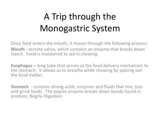
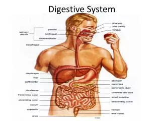
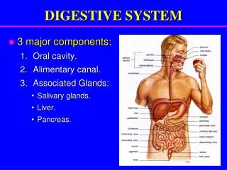
Basic Organization • Mouth • Esophagus • Stomach • Small intestine • Large intestine • Anus
Associated Structures • Pancreas • Liver • Gallbladder • Salivary glands Contribute to small intestinal digestion
Structures in Mouth • Lips • Teeth • Tongue • Salivary glands
Monogastric Teeth • Function: • Mechanically reduce particle size • Increase surface area Four types: • Incisors are used for cutting • Canine (fangs, eye teeth, tusks) are tearing teeth • Premolars and molars (cheek teeth) grind the food
Monogastric Tongue Function: • Comprised of three muscles • Maneuvers food in the mouth • Moves feed to teeth for grinding and to the back of the mouth for swallowing • Can distinguish between feed and toxins by papillae or taste buds
Monogastric Salivary Glands Types of Glands: Zygomatic Parotid Sublingual Mandibular
Functions of Saliva • Moisten feed (salt and water) • Lubrication (aids swallowing) • Starch and(or) lipid digestion (amylase and(or) lipase)
Monogastric Salivary Glands • Flow rate affected by: • Parasympathetic nervous system • Increased tone = Increased flow • Increased flow = Increased dilution • Sympathetic nervous system • Increased tone = Decreased flow • Decreased flow = Increased concentration • Volume of saliva • 1 - 1.5 L/d man and pig • 7 - 10 L/d horse
Monogastric Esophagus • Transport of food from mouth to stomach • Uses peristaltic contractions (wave contractions) • Horse/Pig: • Striated muscles for first 2/3 • Smooth muscles for last 1/3 • In horse, esophagus joins stomach at an oblique angle and cardiac sphincter (the valve between the stomach and esophagus) only allows one-way flow • MOST horses cannot belch out gas or vomit • Dog: • Striated muscles throughout allow GREAT control of digesta movement both directions
Deglutition (Swallowing) • Reflex initiated by presence of food in pharnyx • Propulsion of food to stomach by esophageal peristalsis
Gastric Digestion • Functions • Reservoir for controlled release of digesta to small intestine • Horse has small capacity – requires increased number of smaller sized meals • Mixing food • Mechanical breakdown of feed • Hydrolytic digestion by acid and enzymes • Mainly protein • Kill bacteria • Secrete intrinsic factor: needed for vitamin B12 absorption • Hormone production
Stomach Regions • Esophageal • Non-glandular • Cardiac • Secretes mucus • Fundic • Parietal cells • Chief cells • Pyloric • Mucus
Gastric Pits • Formed by numerous folds in the epithelium • Glands empty into the gastric pit • Many types of glands may empty into one gastric pit
HCl Decreases pH (~2-3) Denatures protein Kills bacteria Activates pepsinogen Mucus Protects lining from acid and enzymes No “autodigestion” Lubricant Pepsinogen Activated form is pepsin Hydrolyzes protein Rennin (abomasum) Clots milk Lipase Some species Stomach Secretions
Gastric Motility and Emptying • Motility aids mixing, mechanical and hydrolytic reduction of feed to chyme • acid pulp • Emptying is stimulated by distension of antral wall and presence of liquid chyme
Control of Gastric Secretions and Gastric Motility • Cephalic phase • Gastric phase • Intestinal phase
Cephalic Phase • Vagal reflex • Parasympathetic innervation • Increases gastric motility, enzyme secretion • Small increase in HCl secretion
Gastric Phase • Local reflex, depends on presence of feed in stomach • Mainly mediated by gastrin • Increases HCl secretion
Intestinal Phase • Stimulated by duodenal distension, pH, osmolarity, nutrients (fat) • Pancreozymin-cholecystokinin (PZ-CCK) is released by the small intestine • Decreases HCl secretion and gastric motility
Gastrointestinal Hormones • Gastrin • Origin: Stomach, Abomasum • Stimulus: Food in stomach • Function: Stimulates HCl & pepsinogen secretion, increases stomach motility • Secretin • Origin: Duodenum • Stimulus: Acid • Function: Stimulates pancreatic secretions. Slows stomach motility and acid production
Gastrointestinal Hormones • Cholecystokinin (CCK) • Origin: Duodenum • Stimulus: Fat & protein in duodenum • Function: Stimulates bile and pancreatic secretions • Also regulates appetite and feed intake • Gastric Inhibitory Protein (GIP) • Origin: Duodenum • Stimulus: Fats and bile • Function: Inhibit stomach motility and secretion of acid and enzymes
Small Intestine • Composed of 3 segments (proximal to distal) • Duodenum • Releases bile and pancreatic secretions • Active site of digestion • Jejunum • Active site of nutrient absorption • Ileum • Active site of nutrient absorption • Most water, vitamins & minerals • Some bacterial presence • Fermentation The pH of the small intestine increases towards 7.0 as food moves from the duodenum to the ileum
Intestinal Epithelial Cell Brush border
Specialized Cells Lining Villi Nutrients Mucus • Absorptive epithelial cell • Contain brush border on lumen/apical side • Brush border: • Enzymes • Nutrient transport molecules • Goblet cell • Secretes mucus
Specialized Cells Lining Villi Anti-microbial compounds • Endocrine cell • Secrete hormones into bloodstream or local cells • Paneth cell • Secretory granules with anti-microbial properties CCK, Secretin, etc.
Small Intestine – Absorptive Surface • Villi • Enterocyte • Brush border • Cell migration from crypts to tips of villus • 2-3 days
Small Intestine - Structure • Lumen • Mucosa • Villi • Crypts • Lacteal • Enterocyte • Brush border
Intestinal Wall Villi Mucosa
Enhanced Surface Area for Increased Nutrient Absorption Intestinal villi
Nutrient Absorption in the Small Intestine • Principal site of absorption of amino acids, vitamins, minerals and lipids • Glucose and other sugars in monogastrics • Generally, most absorption occurs in the proximal (upper) part of the small intestine but some absorption occurs in all segments • Duodenum, jejunum and ileum • Digestion and absorption within SI is rapid • Within 30 minutes of entering SI
Nutrient Absorption • Variety of mechanisms • Diffusion • Facilitated diffusion • Active transport • Pinocytosis or endocytosis • Dependent upon • Solubility of the nutrient (fat vs. water) • Concentration or electrical gradient • Size of the molecule to be absorbed
Diffusion • Water and small lipid molecules pass freely through membrane • Move down concentration gradient to equalize concentrations
Facilitated Diffusion • Carrier loads particle on outside of cell • Carrier releases particle on inside of cell • Reverse Allows equalization of concentrations across membrane
Active Transport • Carrier loads particle on outside of cell • Carrier releases particle on inside of cell • Carrier returns to outside to pick up another particle
Active Transport • Unidirectional movement • Transports nutrients against concentration gradient
Pinocytosis or Endocytosis • Substance contacts cell membrane • Membrane wraps around or engulfs substance into sac • Sac formed separates from the membrane and moves into cell
Secretions Entering SI Secreted from within SI • Intestinal mucus • Brush border enzymes • Pancreatic juices • Produced & stored in pancreas • Bile • Produced in liver • Stored in gallbladder • Horse has no gallbladder • Direct bile secretion into duodenum • Cannot store bile—continuous intake of food Enters from ducts into SI
Intestinal Mucus • Secreted by glands in wall of duodenum • Brunner’s glands • Acts as lubricant and buffer to protect duodenal wall
Bile • Green, viscous liquid • Alkaline ph (neutralize acidic chyme) • Secreted by liver via bile duct to duodenum • Stored in gall bladder (except in horses) • Functions to emulsify fats • Composition • Bile salts (glycocholic and taurocholic acids) • Bile pigments (bilirubin and biliverdin) • Cholesterol • 95% reabsorbed and returned to liver • NOT AN ENZYME
Nutrient Digestion - Lipids Large Lipid Droplet Action of bile salts Lipid emulsion Small Bile salts & pancreatic lipase and colipase Water soluble micelles