DIGESTIVE SYSTEM
DIGESTIVE SYSTEM. GENERAL. 1 st System to Develop “Tube” Within a Tube Contents Remain External Until Absorbed Through Walls Humans are (Anatomical) Omnivores Consists of Alimentary Canal/Digestive Tract and Accessory Organs. DIGESTIVE SYSTEM FUNCTIONS.


DIGESTIVE SYSTEM
E N D
Presentation Transcript
GENERAL • 1st System to Develop • “Tube” Within a Tube • Contents Remain External Until Absorbed Through Walls • Humans are (Anatomical) Omnivores • Consists of Alimentary Canal/Digestive Tract and Accessory Organs
DIGESTIVE SYSTEM FUNCTIONS • Ingestion – Food Enters Through Mouth • Digestion – Breaking Large Molecules into Smaller, Absorbable Molecules • Mechanical (Physical) • Chemical (Enzymatic) • Absorption – Passage of Molecules Through GI Tract Wall into Blood or Lymph • Egestion – Elimination of Non-digestible Substances Through Anal End
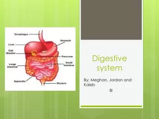
DIGESTIVE SYSTEM ORGANIZATION • Alimentary Canal(Gastrointestinal or Digestive Tract) • Mouth, Pharynx, Esophagus, Stomach, Small Intestine, Large Intestine • Lumen is part of External Environment • Accessory Organs • Salivary Glands • Liver • Gall Bladder • Pancreas
ALIMENTARY CANAL HISTOLOGY • Mucosa • Innermost, Surrounds Lumen • Mucous Membrane • Secretes, Absorbs, Protects • Submucosa • Below Mucosa • C.T., Blood Vessels, Lymphatics, Nervous Tissue
ALIMENTARY CANAL HISTOLOGY continued • Muscularis (Externa) • Smooth Muscle • Inner, Circular, Diameter of Lumen • Outer, Longitudinal, Diameter of Lumen • Propels Substances • Serosa (Visceral Peritoneum) • Outermost • Serous Membrane, Lubricates • Adventitia outside of Ventral Body Cavity
MOVEMENT OF FOOD • Peristalsis - Wave-like Contraction of Circular & Longitudinal Smooth Muscle for Propulsion • ANS • Stimulated by Parasympathetic • Inhibited by Sympathetic
ALIMENTARY CANAL ORGANIZATION • MOUTH • Oral (Buccal) Cavity Functions in Ingestion • Digestion: • Mechanical (Teeth, Tongue) • Chemical (Saliva Begins Carbohydrate Digestion) • Cheeks • Hard Palate (Bony)
MOUTH continued • Soft Palate (Muscular) • Blocks Nasopharynx During Swallowing • Uvula - Dangling end • Tongue • Frenulum Anchors to Floor of Mouth • Skeletal Muscle Tissue covered with Mucosa • Papillae with Taste Buds • Lingual Tonsil & Sublingual Gland
MOUTH continued • Teeth • Vestibule – Space between Teeth & Lips • Two Sets: Deciduous (20) & Permanent (32) • Four Types: • Incisors– Biting • Canines – Tearing & Grasping • Bicuspids & Molars - Grinding
ALIMENTARY CANAL ORGANIZATION continued • PHARYNX • Connects Oral Cavity, Nasal Cavity, Larynx & Esophagus • Passageway for Food, Water, Air • Muscular Walls (Swallowing) • 3 Regions: Nasopharynx, Oropharynx, Laryngopharynx
ALIMENTARY CANAL ORGANIZATION continued • ESOPHAGUS • Collapsible, 10” Tube • Connects Pharynx & Stomach • Lies Posterior to Trachea • Lower Esophageal Sphincter Prevents Stomach Contents from Entering Esophagus • Passes Through Neck Thoracic Cavity Esophageal Hiatus Abdominal Cavity
ALIMENTARY CANAL ORGANIZATION continued • STOMACH • J-Shaped Organ, Inferior to Diaphragm • Functions: • Storage • Digestion (Mechanical & Chemical) • Protein Digestion Begins here • Absorption of Water, Alcohol, Drugs
STOMACH continued • Enters as Bolus – Mixture of Food & Saliva • Leaves as Chyme – Milky Mixture of Partially Digested Food & Gastric Juices
STOMACH continued • 4 Regions: • Cardia – Connects with Esophagus • Body – Principal, Main Part • Fundus – Superior, Bulge, Food Storage • Pylorus – Terminal Part, Pyloric Sphincter • 2 Curves: • Greater Curvature • Lesser Curvature
STOMACH continued • Wall Modifications: • Rugae –Expandable Folds, Accommodate More Food • Oblique Muscle in Muscularis • Gastric Glands in Mucosa • Mucus Cells (Protective Coating) • Parietal Cells (HCl, pH = 2) • Chief Cells (Pepsinogen + HCl Pepsin (Proteolytic)
ALIMENTARY CANAL ORGANIZATION continued • SMALL INTESTINE • Site of Most Chemical Digestion • Site of 90% of Absorption • Monosaccharides Blood • Amino Acids Blood • Fatty Acids & Glycerol Lymph
SMALL INTESTINE continued • 3 Subdivisions: • Duodenum • First 10-12” • Ampulla of Vater Receives Secretions from Liver & Pancreas • Bile through Common Bile Duct • Pancreatic Juice through Pancreatic Duct • Jejunum –Middle Length • Ileum – Last, Joins with Large Intestine at Ileocecal Valve
SMALL INTESTINE continued • Wall Modifications: • All Increase Surface Area for Maximum Absorption • Plicae Circulares – Transverse Folds of Mucosa & Submucosa • Villi – Finger-like Projections of Mucosa, contain Capillaries & Lacteal • Microvilli – Microscopic Folds of Cell Membrane
ALIMENTARY CANAL ORGANIZATION continued • LARGE INTESTINE • Functions: • Secretes Mucus • Absorbs Water, Ions, Vitamins (From Bacterial Metabolism) • Forms & Compacts Feces (Undigested Food, Bacteria, Water)
LARGE INTESTINE continued • 3 Divisions: • Cecum • Blind Pouch • Appendix Attached • Colon - Divided into Ascending, Transverse, Descending, Sigmoid • Rectum – Anal Canal • Internal Anal Sphincter (Smooth) • External Anal Sphincter (Skeletal)
LARGE INTESTINE continued • Wall Modifications: • Taenia Coli – 3 Bands of Longitudinal Muscle from Muscularis of Colon • Haustra – Pouches in Wall of Colon • No Villi
ACCESSORY ORGANS • SALIVARY GLANDS • Exocrine Glands • Ducts Carry Secretions (Saliva) into Mouth • Saliva: Amylase, Mucous, Water, Antibodies, Lysozyme • 3 Pairs: • Parotid (Most Salivary Amylase) • Submandibular • Sublingual
ACCESSORY ORGANS continued • LIVER • Largest Gland in Body (Exocrine) • Produces & Secretes Bile (Fat Emulsifier) • Nutrient Storage & Conversion • Synthesizes Blood Proteins • Detoxification
ACCESSORY ORGANS continued • GALL BLADDER • Muscular Sac • Stores & Concentrates Bile • Receives Bile from Liver via Common Hepatic Duct Cystic Duct • Releases into Duodenum via Cystic Duct Common Bile Duct Ampulla of Vater (Sphincter of Oddi)
ACCESSORY ORGANS continued Bile Release: • Chyme Enters Duodenum • Cholecystokinin (CCK) & Secretin Secreted by Duodenum, Enters Blood • Secretin stimulates liver to produce bile • CCK Stimulates Contractions in Gall Bladder & Relaxation of Sphincter
ACCESSORY ORGANS continued • PANCREAS • Exocrine (& Endocrine) Gland • Secretes Pancreatic Juice (Digestive Enzymes & Buffers) • Leaves Pancreas Through Pancreatic Duct(s), Enters Duodenum Through Ampulla of Vater • Raises pH of Duodenum (pH 8, “Natural Antacid”) via bicarbonate ions