Download
1 / 72
720 likes | 738 Views
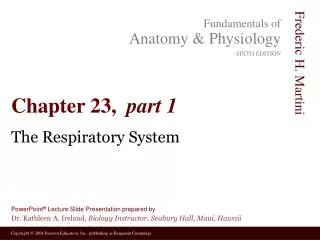
23 PART 1. The Digestive System Pages 676-685, 691-711. Overview of the Digestive System. Organs are divided into two groups Alimentary canal Mouth, pharynx, and esophagus Stomach, small intestine, and large intestine Accessory digestive organs Teeth and tongue

E N D
23 PART 1 The Digestive System Pages 676-685, 691-711
Overview of the Digestive System • Organs are divided into two groups • Alimentary canal • Mouth, pharynx, and esophagus • Stomach, small intestine, and large intestine • Accessory digestive organs • Teeth and tongue • Gallbladder, salivary glands, liver, and pancreas • Accessory organs are connected to the alimentary canal by ducts • Secretions contribute to breakdown of foodstuffs
Parotid gland Mouth (oral cavity) Sublingual gland Salivary glands Tongue Submandibular gland Pharynx Esophagus Stomach Pancreas (Spleen) Liver Gallbladder Transverse colon Duodenum Descending colon Small intestine Jejunum Ascending colon Ileum Cecum Large intestine Sigmoid colon Rectum Vermiform appendix Anus Anal canal The Alimentary Canal and Accessory Digestive Organs Figure 23.1
Digestive Processes • Ingestion—occurs in the mouth • Propulsion—movement of food • Peristalsis—major means of propulsion • Mechanicaldigestion—prepares food for chemical digestion • Chewing, churning food in stomach, segmentation • Segmentation is rhythmic local constrictions of intestine
Digestive Processes • Chemical digestion—complex molecules broken down to chemical components • Mouth • Stomach • Small intestine • Absorption—transport of digested nutrients • Defecation—elimination of indigestible substances as feces
Ingestion Food Mechanical digestion Pharynx Esophagus • Chewing (mouth) • Churning (stomach) Propulsion • Segmentation (small intestine) • Swallowing (oropharynx) Chemical digestion • Peristalsis (esophagus, stomach, small intestine, large intestine) Stomach Absorption Lymph vessel Small intestine Blood vessel Large intestine Mainly H2O Feces Anus Defecation Digestive Processes Figure 23.5
Peristalsis • Major means of propulsion • Adjacent segments of the alimentary canal relax and contract Figure 23.6
Segmentation • Rhythmic local contractions of the intestine • Mixes food with digestive juices Figure 23.6
The Peritoneal Cavity and Peritoneum • Peritoneum—a serous membrane • Visceral peritoneum—surrounds digestive organs • Parietal peritoneum—lines the body wall • Peritoneal cavity—a slit-like potential space
The Peritoneal Cavity and Peritoneum • Mesentery—a double layer of peritoneum • Holds organs in place • Sites of fat storage • Provides a route for circulatory vessels and nerves
Alimentary canal organ Abdominopelvic cavity Alimentary canal organ in a retroperitoneal position Liver Ventral mesentery Alimentary canal organ Parietal peritoneum Visceral peritoneum Peritoneal cavity Dorsal mesentery Mesentery resorbed and lost Vertebra (a) Schematic cross sections of abdominal cavity illustrating the peritonea and mesenteries (c) Some organs lose their mesentery and become retroperitoneal during development. Visceral peritoneum Anterior Falciform ligament Peritoneal cavity (with serous fluid) Liver Stomach Parietal peritoneum Kidney (retroperitoneal) Wall of body trunk Posterior (b) Illustration of the peritonea in a cross section through the superior abdomen, inferior view The Peritoneal Cavity and Peritoneum
The Peritoneal Cavity and Peritoneum • Retroperitoneal organs • Behind the peritoneum • Peritoneal organs • Digestive organs that keep their mesentery
Falciform ligament Liver Gallbladder Spleen Stomach Ligamentum teres Greater omentum Small intestine Cecum (a) Mesenteries • Superficial view of abdominal organs Figure 23.4
Liver Gallbladder Lesser omentum Stomach Duodenum Transverse colon Small intestine Cecum Urinary bladder (b) Mesenteries • Lesser omentum attaches to lesser curvature of stomach Figure 23.4
Greater omentum Transverse colon Transverse mesocolon Descending colon Jejunum Mesentery Sigmoid mesocolon Sigmoid colon Ileum (c) Mesenteries • Greater omentum—a “fatty apron” of peritoneum • Greater omentum and transverse colon reflected Figure 23.4
Liver Lesser omentum Pancreas Stomach Duodenum Transverse mesocolon Transverse colon Mesentery Greater omentum Jejunum Ileum Visceral peritoneum Parietal peritoneum Urinary bladder Rectum (d) Mesenteries • Sagittal section through the abdominopelvic cavity • Mesenteries attach to posterior abdominal wall
Secondarily Retroperitoneal Organs • Initially formed within peritoneum • Become retroperitoneal • Fuse to posterior abdominal wall
Summary of Intraperitoneal and Secondarily Retroperitoneal Organs Table 23.1
Histology of the Alimentary Canal Wall • Same four layers from esophagus to anus • The mucosa—innermost layer • Consists of • Epithelium • Lamina propria • Muscularis mucosae • The submucosa—external to the mucosa • Contains blood and lymphatic vessels, nerve fibers
Intrinsic nerve plexuses Myenteric nerve plexus Submucosal nerve plexus Glands in submucosa Mucosa Epithelium Lamina propria Muscularis mucosae Submucosa Muscularis externa Longitudinal muscle Circular muscle Serosa Epithelium Connective tissue Nerve Lumen Artery Gland in mucosa Vein Duct of gland outside alimentary canal Mucosa-associated lymphoid tissue Lymphatic vessel Mesentery (a) Longitudinal and cross-sectional views through the small intestine Histology of the Alimentary Canal Figure 23.7a
Histology of the Alimentary Canal Wall • The muscularis externa—external to the submucosa • Two layers • Circular muscularis—inner layer • Longitudinal muscularis—outer layer • The serosa—the outermost layer • Is the visceral peritoneum
Mucosa Submucosa Muscularis externa Serosa (b) Light micrograph cross section through the small intestine (30) Histology of the Alimentary Canal Figure 23.7b
Smooth Muscle • Primarily found in walls of viscera • Fibers elongated • Have one centrally located nucleus • Grouped into sheets • Longitudinal layer—parallel to long axis of organ • Circular layer—deeper layer, fibers run around circumference of organ
Longitudinal layer of smooth muscle (shows smooth muscle fibers in cross section, 145) Small intestine Mucosa (b) Cross section of the intestine showing the smooth muscle layers (one circular and the other longitudinal) running at right angles to each other Circular layer of smooth muscle (shows longitudinal views of smooth muscle fibers, 145) (a) Location and plane of section shown in (b) Smooth Muscle Figure 23.8a, b
The Salivary Glands Produce saliva Parotid glands Parotid duct—parallel to zygomatic arch Submandibular glands Lie along medial surface of mandible Sublingual glands Lie in floor of oral cavity
Figure 23.15 The major salivary glands. Tongue Teeth Parotidgland Ducts ofsublingualgland Parotid duct Masseter muscle Frenulumof tongue Body of mandible(cut) Sublingualgland Posterior belly ofdigastric muscle Mylohyoidmuscle (cut) Submandibularduct Anterior belly ofdigastric muscle Submandibulargland
The Pharynx • Oropharynx and laryngopharynx • Passages for air and food • Lined with stratified squamous epithelium • External muscle layer • Consists of superior, middle, and inferior pharyngeal constrictors
The Esophagus • Gross anatomy—muscular tube • Begins as a continuation of the pharynx • Joins the stomach inferior to the diaphragm • Cardiac sphincter—closes lumen to prevent stomach acid from entering esophagus
Parotid gland Mouth (oral cavity) Sublingual gland Salivary glands Tongue Submandibular gland Pharynx Esophagus Stomach Pancreas (Spleen) Liver Gallbladder Transverse colon Duodenum Descending colon Small intestine Jejunum Ascending colon Ileum Cecum Large intestine Sigmoid colon Rectum Vermiform appendix Anus Anal canal The Alimentary Canal and Accessory Digestive Organs Figure 23.1
Xiphoid process of sternum Foramen for inferior vena cava Foramen for esophagus-esophageal hiatus Costal cartilage Central tendon of diaphragm Diaphragm Foramen for aorta Lumbar vertebra 12th rib Quadratus lumborum Psoas major (b) Diaphragm Figure 11.13b
Cardia Fundus Esophagus Serosa Muscularis externa Longitudinal layer Circular layer Body Oblique layer Lumen Lesser curvature Rugae of mucosa Greater curvature Pyloric canal Pyloric antrum Duodenum Pyloric sphincter (valve) at pylorus (a) Figure 23.17a
The Esophagus • Microscopic anatomy • Epithelium is stratified squamous epithelium • When empty, mucosa and submucosa in longitudinal folds • Mucous glands—extends from submucosa to lumen • Muscularis externa • Skeletal muscle first third of length • Adventitia—most external layer
Figure 23.16 Microscopic structure of the esophagus. Mucosa(stratified squamousepithelium) Esophagus-stomachjunction Submucosa(areolar connective tissue) Lumen Muscularis externa Simple columnarepithelium ofstomach Circular layer Longitudinal layer Adventitia (fibrousconnective tissue) Cross section throughesophagus (3) Esophagus-stomach junction,longitudinal section (85)
The Stomach • Site where food is churned into chyme • Secretion of pepsin begins protein digestion • Functions under acidic conditions • Food remains in stomach approximately 4 hours • Regions of the stomach • Cardiac region • Fundus • Body • Pyloric region
Cardia Fundus Esophagus Serosa Muscularis externa Longitudinal layer Circular layer Body Oblique layer Lumen Lesser curvature Rugae of mucosa Greater curvature Pyloric canal Pyloric antrum Duodenum Pyloric sphincter (valve) at pylorus (a) The Stomach Figure 23.17a
Fundus Liver (cut) Body Spleen Lesser curvature Greater curvature (b) The Stomach Figure 23.17b
Microscopic Anatomy of the Stomach • Muscularis has three layers • Circular and longitudinal layers and oblique layer • Epithelium is simple columnar epithelium • Mucosa dotted with gastric pits • Gastric glands—deep to gastric pits
Microscopic Anatomy of the Stomach • Gastric glands of fundus and body • Mucous neck cells • Secrete a special mucus • Parietal (oxyntic) cells • Secrete hydrochloric acid and gastric intrinsic factor • Chief (zymogenic) cells • Secrete pepsinogen • Pepsinogen is activated to pepsin when it encounters acid in the gastric glands
Gastric pits Surface epithelium (mucous cells) Surface epithelium Gastric pit Mucous neck cells Mucosa Parietal cell Gastric gland Lamina propria Muscularis mucosae Submucosa (contains submucosal plexus) Chief cell Oblique layer Circular layer Muscularis externa (contains myenteric plexus) Longitudinal layer Enteroendocrine cell Serosa (b) Enlarged view of gastric pits and gastric glands Stomach wall (a) Layers of the stomach wall, longitudinal section Pepsinogen Pepsin HCl Mitochondria Parietal cell Chief cell Enteroendocrine cell (c) Location of the HCl-producing parietal cells and pepsin-secreting chief cells in a gastric gland The Stomach—Microscopic Anatomy Figure 23.18
Figure 23.18 Microscopic anatomy of the stomach. Gastric pits Surface epithelium(mucous cells) Gastricpit Surfaceepithelium Mucous neck cells Parietal cell Gastricgland Mucosa Lamina propria Chief cell Muscularismucosae Submucosa(containssubmucosalplexus) Obliquelayer Enteroendocrine cell Pepsinogen Pepsin HCl Enlarged view ofgastric pits andgastric glands Circularlayer Muscularisexterna(containsmyentericplexus) Longitudinallayer Mitochondria Parietal cell Stomach wall Serosa Layers of the stomach wall, longitudinal section Chief cell Mucus-secretingcells Gastric pits Enteroendocrinecell Surface mucous cell Mucous neck cells Location of the HCl-producing parietalcells and pepsin-secreting chief cells ina gastric gland HCl-secreting parietal cells Gastricgland Enzyme-secreting chief cells Muscularismucosae Micrograph of the stomach mucosa, view similar to part (b) (110)
The Small Intestine—Gross Anatomy • Longest portion of the alimentary canal • Site of most enzymatic digestion and absorption • Three subdivisions • Duodenum • Jejunum • Ileum
The Duodenum • Receives digestive enzymes and bile • Main pancreatic duct and common bile duct enter duodenum • Sphincters control entry of bile and pancreatic juices
Right and left hepatic ducts of liver Common hepatic duct Cystic duct Bile duct and sphincter Accessory pancreatic duct Mucosa with folds Tail of pancreas Pancreas Gallbladder Jejunum Major duodenal papilla Main pancreatic duct and sphincter Hepatopancreatic ampulla and sphincter Duodenum Head of pancreas The Duodenum and Related Organs Figure 23.19
The Small Intestine—Microscopic Anatomy • Modifications for absorption • Circular folds (plicae circulares) • Transverse ridges of mucosa and submucosa • Villi • Finger-like projections of the mucosa • Covered with simple columnar epithelium • Microvilli • Further increase surface area for absorption
Histology of the Intestinal Wall • Absorptive cells • Uptake digested nutrients • Goblet cells • Secrete mucus that lubricates chyme • Enteroendocrine cells • Secrete hormones • Intestinal crypts • Epithelial cells secrete intestinal juice
Microvilli (brush border) Absorptive cells Lacteal Goblet cell Blood capillaries Vilus Absorptive cells Mucosa associated lymphoid tissue Goblet cells Villi Enteroendocrine cells Intestinal crypt Venule Muscularis mucosae Lymphatic vessel Duodenal gland Submucosa (b) (c) Intestinal crypt The Small Intestine—Structural Features Vein carrying blood to hepatic portal vessel Muscle layers Lumen Circular folds Villi (a) Figure 23.20
The Large Intestine • Digested residue contains few nutrients • Small amount of digestion by bacteria • Main functions • Absorb water and electrolytes
Gross Anatomy of Large Intestine • Subdivided into • Cecum, vermiform appendix, colon, rectum, anal canal • Special features of large intestine • Teniae coli • Thickening of longitudinal muscularis • Haustra • Puckering created by teniae coli • Epiploic appendages • Fat-filled pouches of visceral peritoneum