Chapter 8 Skeletal System
Chapter 8 Skeletal System. Introduction. Skeletal tissues form bones—the organs of the skeletal system The relationship of bones to each other and to other body structures provides a basis for understanding the function of other organ systems

Chapter 8 Skeletal System
E N D
Presentation Transcript
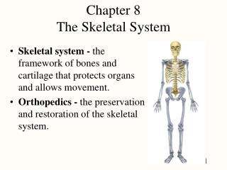
Introduction • Skeletal tissues form bones—the organs of the skeletal system • The relationship of bones to each other and to other body structures provides a basis for understanding the function of other organ systems • The adult skeleton is composed of 206 separate bones
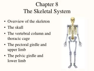
Divisions of Skeleton • Axial skeleton—the 80 bones of the head, neck, and torso; composed of 74 bones that form the upright axis of the body and six tiny middle ear bones
Divisions of Skeleton • Appendicular skeleton—the 126 bones that form the appendages to the axial skeleton; the upper and lower extremities
Axial Skeleton • Skull—made up of 28 bones in two major divisions: cranial bones and facial bones (Figures 8-2 to 8-7; Table 8-3)
Cranial bones • Frontal bone • Forms the forehead and anterior part of the top of the cranium • Contains the frontal sinuses • Forms the upper portion of the orbits • Forms the coronal suture with the two parietal bones
Cranial bones • Parietal bones • Form the bulging top of the cranium • Form several sutures: lambdoidal suture with occipital bone; squamous suture with temporal bone and part of sphenoid; and coronal suture with frontal bone
Cranial bones • Temporal bones • Form the lower sides of the cranium and part of the cranial floor • Contain the inner and middle ears
Cranial bones • Occipital bone • Forms the lower, posterior part of the skull • Forms immovable joints with three other cranial bones and a movable joint with the first cervical vertebra
Cranial bones • Sphenoid bone • A bat-shaped bone located in the central portion of the cranial floor • Anchors the frontal, parietal, occipital, and ethmoid bones and forms part of the lateral wall of the cranium and part of the floor of each orbit (Figure 8-7) • Contains the sphenoid sinuses
Cranial bones • Ethmoid bone • A complicated, irregular bone that lies anterior to the sphenoid and posterior to the nasal bones • Forms the anterior cranial floor, medial orbit walls, upper parts of the nasal septum, and sidewalls of the nasal cavity • The cribriform plate is located in the ethmoid
Facial bones • Maxilla (upper jaw) • Two maxillae form the keystone of the face • Maxillae articulate with each other and with nasal, zygomatic, inferior concha, and palatine bones • Forms parts of the orbital floors, roof of the mouth, and floor and sidewalls of the nose • Contains maxillary sinuses
Facial bones • Mandible (lower jaw) • Largest, strongest bone of the face • Forms the only movable joint of the skull with the temporal bone
Facial bones • Zygomatic bone • Shapes the cheek and forms the outer margin of the orbit • Forms the zygomatic arch with the zygomatic process of the temporal bones
Facial bones • Nasal bone • Both nasal bones form the upper part of the bridge of the nose, whereas cartilage forms the lower part • Articulates with the ethmoid bone, nasal septum, frontal bone, maxillae, and the other nasal bone
Facial bones • Lacrimal bone • Paper-thin bone that lies just posterior and lateral to each nasal bone • Forms the nasal cavity and medial wall of the orbit • Contains groove for the nasolacrimal (tear) duct • Articulates with the maxilla and the frontal and ethmoid bones
Facial bones • Palatine bone • Two bones form the posterior part of the hard palate • Vertical portion forms the lateral wall of the posterior part of each nasal cavity • Articulates with the maxillae and the sphenoid bone
Axial Skeleton • Facial bones (cont.) • Inferior nasal conchae (turbinates) • Form lower edge projecting into the nasal cavity and form the nasal meati • Articulate with ethmoid, lacrimal, maxillary, and palatine bones • Vomer bone (Figure 8-8, G) • Forms posterior portion of the nasal septum • Articulates with the sphenoid, ethmoid, and palatine bones and maxillae
Axial Skeleton • Eye orbits • Right and left eye orbits • Contain eyes, associated eye muscles, lacrimal apparatus, blood vessels, and nerves • Thin and fragile orbital walls separate orbital structures from cranial and nasal cavities and paranasal sinuses • Traumatic injuries may result in “blowout fractures” • “Raccoon eyes”—clinical sign of blowout fracture
Fetal skull • Characterized by unique anatomic features not seen in adult skull • Fontanels or “soft spots” (4) allow skull to “mold” during birth process and permit rapid growth of brain
Fetal skull • Permits differential growth or appearance of skull components over time • Face—smaller proportion of total cranium at birth (1/8) than in adult (1/2) • Head at birth is ¼ total body height; at maturity is about 1/8 body height • Sutures appear with skeletal maturity • Paranasal sinuses—change in size and placement with skeletal maturity • Appearance of deciduous and, later, permanent teeth
Hyoid bone • U-shaped bone located just above the larynx and below the mandible • Suspended from the styloid processes of the temporal bone • Only bone in the body that articulates with no other bones
Vertebral column • Forms the flexible longitudinal axis of the skeleton • Consists of 24 vertebrae plus the sacrum and coccyx • Segments of the vertebral column: • Cervical vertebrae, 7 • Thoracic vertebrae, 12 • Lumbar vertebrae, 5 • Sacrum—in adult, results from fusion of five separate vertebrae • Coccyx—in adult, results from fusion of four or five separate vertebrae
Vertebral column • Characteristics of the vertebrae • All vertebrae, except the first, have a flat, rounded body anteriorly and centrally, a spinous process posteriorly, and two transverse processes laterally • All but the sacrum and coccyx have vertebral foramen • Second cervical vertebra has upward projection, the dens, to allow rotation of the head • Seventh cervical vertebra has long, blunt spinous process • Each thoracic vertebra has articular facets for the ribs
Vertebral column • Vertebral column as a whole articulates with the head, ribs, and iliac bones • Individual vertebrae articulate with each other in joints between their bodies and between their articular processes
Sternum • Dagger-shaped bone in the middle of the anterior chest wall made up of three parts: • Manubrium—the upper, handle part • Body—the middle, blade part • Xiphoid process—the blunt cartilaginous lower tip, which ossifies during adult life
Sternum • Manubrium articulates with the clavicle and first rib • Next nine ribs join the body of the sternum, either directly or indirectly, by means of the costal cartilage
Ribs • Twelve pairs of ribs, with the vertebral column and sternum, form the thorax • Each rib articulates with the body and transverse process of its corresponding thoracic vertebra • Ribs 2 through 9 articulate with the body of the vertebra above
Ribs • From its vertebral attachment, each rib curves outward, then forward and downward • Rib attachment to the sternum: • Ribs 1 through 8 join a costal cartilage that attaches it to the sternum • Costal cartilage of ribs 8 through 10 joins the cartilage of the rib above to be indirectly attached to the sternum • Ribs 11 and 12 are floating ribs, because they do not attach even indirectly to the sternum
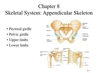
Appendicular Skeleton • Upper extremity • Consists of the bones of the shoulder girdle, upper arm, lower arm, wrist, and hand • Shoulder girdle • Made up of scapula and clavicle • Clavicle forms only bony joint with trunk, the sternoclavicular joint • At its distal end, clavicle articulates with the acromion process of the scapula
Appendicular Skeleton • Upper extremity (cont.) • Humerus • The long bone of the upper arm • Articulates proximally with the glenoid fossa of the scapula and distally with the radius and ulna
Appendicular Skeleton • Upper extremity (cont.) • Ulna • Long bone found on little finger side of forearm • Articulates proximally with humerus and radius and distally with a fibrocartilaginous disk
Appendicular Skeleton • Upper extremity (cont.) • Radius • Long bone found on thumb side of forearm • Articulates proximally with capitulum of humerus and radial notch of ulna; articulates distally with scaphoid and lunate carpals and with head of ulna
Appendicular Skeleton • Upper extremity (cont.) • Carpal bones • Eight small bones that form wrist • Carpals are bound closely and firmly by ligaments and form two rows of four carpals each • Proximal row is made up of pisiform, triquetrum, lunate, and scaphoid • Distal row is made up of hamate, capitate, trapezoid, and trapezium • The joints between radius and carpals allow wrist and hand movements
Appendicular Skeleton • Upper extremity (cont.) • Metacarpal bones • Form framework of hand • Thumb metacarpal forms the most freely movable joint with the carpals • Heads of metacarpals (knuckles) articulate with phalanges
Appendicular Skeleton • Lower extremity • Consists of the bones of hip, thigh, lower leg, ankle, and foot
Appendicular Skeleton • Lower extremity • Pelvic girdle is made up of the sacrum and the two coxal bones, bound tightly by strong ligaments • A stable circular base that supports the trunk and attaches the lower extremities to it • Each coxal bone is made up of three bones that fuse together: • Ilium—largest and uppermost • Ischium—strongest and lowermost • Pubis—anteriormost
Appendicular Skeleton • Lower extremity (cont.) • Femur—longest and heaviest bone in the body • Patella—largest sesamoid bone in the body • Tibia • The larger, stronger, and more medially and superficially located of the two leg bones • Articulates proximally with the femur to form the knee joint • Articulates distally with the fibula and the talus
Appendicular Skeleton • Lower extremity (cont.) • Fibula • The smaller, more laterally and deeply placed of two leg bones • Articulates with tibia
Appendicular Skeleton • Lower extremity (cont.) • Foot (Figures 8-24 and 8-25) • Structure is similar to that of the hand, with adaptations for supporting weight • Foot bones are held together to form spring arches • Medial longitudinal arch is made up of calcaneus, talus, navicular, cuneiforms, and medial three metatarsals • Lateral longitudinal arch is made up of calcaneus, cuboid, and fourth and fifth metatarsals
Skeletal Differences in Men and Women • Male skeleton is larger and heavier than female skeleton • Pelvic differences • Male pelvis—deep and funnel-shaped with a narrow pubic arch • Female pelvis—shallow, broad, and flaring with a wider pubic arch
Cycle of Life: The Aging Skeleton • Aging changes begin at fertilization and continue over a lifetime • Changes can be positive or negative • Normal bone development is a skeletal aging process • Intramembranous ossification • Endochondral ossification • Appearance of ossification centers and closure of epiphyseal plates can be used to estimate potential growth and height
Cycle of Life: The Aging Skeleton • Characteristics of bone during age • Bone produced early in life is properly calcified but not brittle • Osteoblastic activity during early periods of bone remodeling results in deposition of more bone than is resorbed • Prior to puberty results in growth of bones • After puberty and until early thirties, replaced bone is stronger
Cycle of Life: The Aging Skeleton • Negative outcomes of skeletal aging begin between 30 and 40 years of age • Decrease in osteoblast numbers with production of lower quality matrix • Increase in osteoclast numbers and activity with increased bone loss • Mature osteocytes coalesce and shrink, producing a honeycomb of tiny holes in the compact bone
Cycle of Life: The Aging Skeleton • Negative outcomes (cont.) • Skeleton as a whole loses strength, and fracture risk increases • Decrease in number of trabeculae in spongy bone in vertebral bodies and other bones results in “spontaneous” as well as compression fractures • Overall height decreases beginning at about age 35 • Osteoporosis is a common and very serious bone disease in old age
Mechanisms of Disease—Bone Fractures • Fracture defined as partial or complete break in continuity of a bone • Mechanical stress and traumatic injury are most common causes • Pathological or spontaneous fractures occur in absence of trauma • Stress fractures may not be apparent in clinical examination or standard x-ray images but can be seen in bone scans • Bone damage is microscopic • Caused by repetitive trauma (e.g., marathon runners)