Download
1 / 51
530 likes | 889 Views
Chapter 8: Skeletal Muscle. EXERCISE PHYSIOLOGY Theory and Application to Fitness and Performance, 6 th edition Scott K. Powers & Edward T. Howley. Objectives. Draw & label the microstructure of skeletal muscle Outline the steps leading to muscle shortening

E N D
Chapter 8:Skeletal Muscle EXERCISE PHYSIOLOGY Theory and Application to Fitness and Performance, 6th edition Scott K. Powers & Edward T. Howley
Objectives • Draw & label the microstructure of skeletal muscle • Outline the steps leading to muscle shortening • Define the concentric and isometric • Discuss: twitch, summation & tetanus • Discus the major biochemical and mechanical properties of skeletal muscle fiber types
Objectives • Discuss the relationship between skeletal muscle fibers types and performance • List & discuss those factors that regulate the amount of force exerted during muscular contraction • Graph the relationship between movement velocity and the amount of force exerted during muscular contraction • Discuss structure & function of muscle spindle • Describe the function of a Golgi tendon organ
Skeletal Muscle • Human body contains over 400 skeletal muscles • 40-50% of total body weight • Functions of skeletal muscle • Force production for locomotion and breathing • Force production for postural support • Heat production during cold stress
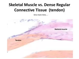
Connective Tissue Covering Skeletal Muscle • Epimysium • Surrounds entire muscle • Perimysium • Surrounds bundles of muscle fibers • Fascicles • Endomysium • Surrounds individual muscle fibers
Microstructure of Skeletal Muscle • Sarcolemma: Muscle cell membrane • Myofibrils Threadlike strands within muscle fibers • Actin (thin filament) • Myosin (thick filament) • Sarcomere • Z-line, M-line, H-zone, A-band & I-band
Microstructure of Skeletal Muscle • Within the sarcoplasm • Sarcoplasmic reticulum • Storage sites for calcium • Transverse tubules • Terminal cisternae • Mitochondria
Within the Sarcoplasm Fig 8.3
The Neuromuscular Junction • Where motor neuron meets the muscle fiber • Motor end plate: pocket formed around motor neuron by sarcolemma • Neuromuscular cleft: short gap • Ach is released from the motor neuron • Causes an end-plate potential (EPP) • Depolarization of muscle fiber
Neuromuscular Junction Fig 8.4
Muscular Contraction • The sliding filament model • Muscle shortening occurs due to the movement of the actin filament over the myosin filament • Formation of cross-bridges between actin and myosin filaments “Power stroke” • 1 power stroke only shorten muscle 1% • Reduction in the distance between Z-lines of the sarcomere
The Sliding Filament Model Fig 8.5
Actin & Myosin Relationship • Actin • Actin-binding site • Troponin with calcium binding site • Tropomyosin • Myosin • Myosin head • Myosin tais
Actin & Myosin Relationship Fig 8.6
Energy for Muscle Contraction • ATP is required for muscle contraction • Myosin ATPase breaks down ATP as fiber contracts • Sources of ATP • Phosphocreatine (PC) • Glycolysis • Oxidative phosphorylation
Excitation-Contraction Coupling • Depolarization of motor end plate (excitation) is coupled to muscular contraction • Nerve impulse travels down T-tubules and causes release of Ca++ from SR • Ca++ binds to troponin and causes position change in tropomyosin, exposing active sites on actin • Permits strong binding state between actin and myosin and contraction occurs
Excitation-Contraction Coupling Fig 8.9
Properties of Muscle Fiber Types • Biochemical properties • Oxidative capacity • Type of ATPase • Contractile properties • Maximal force production • Speed of contraction • Muscle fiber efficiency
Fast fibers Type IIx fibers Fast-twitch fibers Fast-glycolytic fibers Type IIa fibers Intermediate fibers Fast-oxidative glycolytic fibers Slow fibers Type I fibers Slow-twitch fibers Slow-oxidative fibers Individual Fiber Types
Muscle Fiber Types Fast FibersSlow fibers Characteristic Type IIx Type IIa Type I Number of mitochondria Low High/mod High Resistance to fatigue Low High/mod High Predominant energy system Anaerobic Combination Aerobic ATPase Highest High Low Vmax (speed of shortening) Highest Intermediate Low Efficiency Low Moderate High Specific tension High High Moderate
Type IIx Comparison of Maximal Shortening Velocities Between Fiber Types Fig 8.11
Histochemical Staining of Fiber Type Type IIa Type IIx Type I Fig 8.12
+ 1 2 3 4 5 Type I Type IIA Type IIx 5 - Biceps 4 – Quads 3 – Gastroc 2 – Soleus 1 – Marker _ Fiber Typing • Gel electrophoresis: myosin isoforms • different weight move different distances Table 10.2
Fiber Typing • Immunohistochemical: • Four serial slices of muscle tissue • antibody attach to certain myosin isoforms
Muscle Tissue: Rat Diaphragm Type 1 fibers - dark antibody: BA-D5 Type 2b fiber - dark Antibody: BF-F3 Type 2x fibers - light/white antibody: BF-35 Type 2a fibers - dark antibody: SC-71
Fiber Types and Performance • Power athletes • Sprinters • Possess high percentage of fast fibers • Endurance athletes • Distance runners • Have high percentage of slow fibers • Others • Weight lifters and nonathletes • Have about 50% slow and 50% fast fibers
Alteration of Fiber Type by Training • Endurance and resistance training • Cannot change fast fibers to slow fibers • Can result in shift from Type IIx to IIa fibers • Toward more oxidative properties
Age-Related Changes in Skeletal Muscle • Aging is associated with a loss of muscle mass • Rate increases after 50 years of age • Regular exercise training can improve strength and endurance • Cannot completely eliminate the age-related loss in muscle mass
Types of Muscle Contraction • Isometric • Muscle exerts force without changing length • Pulling against immovable object • Postural muscles • Isotonic (dynamic) • Concentric • Muscle shortens during force production • Eccentric • Muscle produces force but length increases
Isotonic and Isometric Contractions Fig 8.14
Speed of Muscle Contraction and Relaxation • Muscle twitch • Contraction as the result of a single stimulus • Latent period • Lasting only ~5 ms • Contraction • Tension is developed • 40 ms • Relaxation • 50 ms
Muscle Twitch Fig 8.15
Force Regulation in Muscle • Types and number of motor units recruited • More motor units = greater force • Fast motor units = greater force • Increasing stimulus strength recruits more & faster/stronger motor units • Initial muscle length • “Ideal” length for force generation • Nature of the motor units neural stimulation • Frequency of stimulation • Simple twitch, summation, and tetanus
Relationship Between Stimulus Frequency and Force Generation Fig 8.16
Length-Tension Relationship Fig 8.17
Force-Velocity Relationship • At any absolute force the speed of movement is greater in muscle with higher percent of fast-twitch fibers • The maximum velocity of shortening is greatest at the lowest force • True for both slow and fast-twitch fibers
Force-Velocity Relationship Fig 8.19
Force-Power Relationship • At any given velocity of movement the power generated is greater in a muscle with a higher percent of fast-twitch fibers • The peak power increases with velocity up to movement speed of 200-300 degrees•second-1 • Force decreases with increasing movement speed beyond this velocity
Force-Power Relationship • At any given velocity of movement the power generated is greater in a muscle with a higher percent of fast-twitch fibers • The peak power increases with velocity up to movement speed of 200-300 degrees/sec • Force decreases with increasing movement speed beyond this velocity
Force-Power Relationship Fig 8.20
Receptors in Muscle • Muscle spindle • Changes in muscle length • Rate of change in muscle length • Intrafusal fiber contains actin & myosin, and therefore has the ability to shorten • Gamma motor neuron stimulate muscle spindle to shorten • Stretch reflex • Stretch on muscle causes reflex contraction
Muscle Spindle Fig 8.21
Receptors in Muscle • Golgi tendon organ (GTO) • Monitor tension developed in muscle • Prevents damage during excessive force generation • Stimulation results in reflex relaxation of muscle
Golgi Tendon Organ Fig 8.22