Chapter 8 The Skeletal System
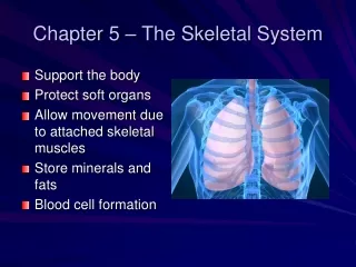
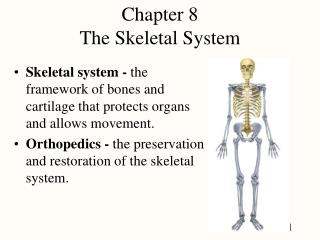
Chapter 8 The Skeletal System. Skeletal system - the framework of bones and cartilage that protects organs and allows movement. Orthopedics - the preservation and restoration of the skeletal system. Overview of the Skeleton. Regions of the skeleton 1. axial skeleton forms the central axis

Chapter 8 The Skeletal System
E N D
Presentation Transcript
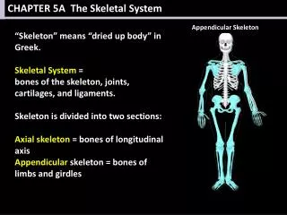
Chapter 8The Skeletal System • Skeletal system - the framework of bones and cartilage that protects organs and allows movement. • Orthopedics - the preservation and restoration of the skeletal system.
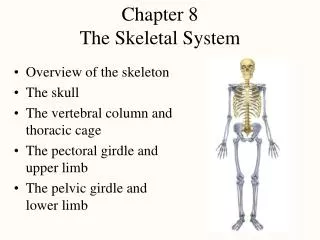
Overview of the Skeleton • Regions of the skeleton • 1. axial skeleton forms the central axis • skull, vertebral column, ribs, sternum and sacrum • 2. appendicular skeleton includes the limbs & girdles • Number of bones • 206 in typical adult skeleton
Surface Features of Bones • 1. joint formation • 2. muscle attachment • 3. passage of nerves and blood vessels these are foramen.
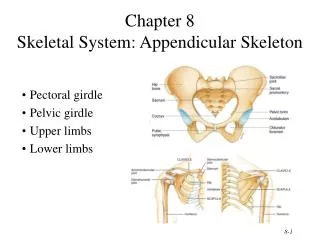
Axial & Appendicular Skeleton • Axial skeleton (yellow) • skull, vertebrae, ribs, sacrum & hyoid even auditory ossicles • Appendicular skeleton (blue) • pectoral girdle • upper extremity • pelvic girdle • lower extremity
The Skull • 22 bones joined together by sutures • Cranial bones surround cranial cavity • 8 bones in contact with meninges • Frontal, Sphenoid, Occipital, Ethmoid, (2) Temporal, (2) Parietal • Facial bones support teeth & form nasal cavity & orbit • 14 bones with no direct contact with brain or meninges • (2) Nasal bones, (2) Maxillae, (2) Zygomatic, (2) Lacrimal, (2)Palatine, (2) Inf. Nasal conchae, (1) Vomer, (1) Mandible
Sutures • Sutures are movable joints* found only between skull bones: • 1. coronal • 2. sagittal • 3. lambdoid • 4. squamous *Bone of contention, clinically skull bones do move. The anatomists say they don’t. Why?
Cranial Fossa • 3 basins that comprise the cranial floor or base • anterior fossa holds the frontal lobe of the brain • middle fossa holds the temporal lobes of the brain • posterior fossa contains the cerebellum
Frontal Bone Parietal Bone A Paired Bone
Temporal BoneBoxed Ear Bone • 3 parts: • squamous part • zygomatic process • Petrous part • Ear • styloid process for muscle attachment • mastoid process • looks like a _______
Petrous Portion of Temporal Bone • Houses the EAR • Petrous means _______
Occipital BoneThe Basic Bone • Rear & much of base of skull • Foramen magnum holds spinal cord • Skull rests on atlas at occipital condyles
Sphenoid BoneTHE Skull bone. • Lesser wing • Greater wing • Medial and lateral pterygoid processes
Sphenoid BoneTHE Keystone of the skull. • Body of the sphenoid • sella turcica (Turkish Saddle) • houses pituitary gland • Lesser wing • Optic foramen • Greater wing -- 3 foramen • foramen rotundum & ovale for brs. trigeminal nerve • foramen spinosum for meningeal artery
Ethmoid BoneThe Stinky Bone • Found between the orbital cavities • Forms lateral walls and roof of nasal cavity • Crista galli is a structure inside the cranium • Cribriform plate has holes for CN 1 • Ethmoid air cells form ethmoid sinus • Perpendicular plate forms the superior part of nasal septum • Concha or turbinates on lateral wall
Ethmoid BoneThe Smelly Bone • Superior & middle concha • Perpendicular plate of nasal septum • CN 1 goes to the nose
2 Maxillary Bones Maxium Face Bone • Forms upper jaw • Hold upper teeth • Forms inferomedial wall of orbit • Forms anterior 2/3’sof hard palate • cleft palate
Zygomatic BonesCheek Bone • Forms cheekbones and lateral orbit • Zygomatic arch is formed from: • temporal process of zygomatic bone • zygomatic process of temporal bone
2 Nasal Bones • Forms bridge of nose and supports cartilages of nose • Often fractured by trauma to the nose 2 Lacrimal Bones
2 Inferior Nasal Conchae • A separate bone • Not a part of the ethmoid like the superior & middle concha
Vomer • Inferior half of the nasal septum • Supports cartilage of nasal septum
Palatine Bones • L-shaped bone • Posterior 1/3 of the hard palate • Part of lateral nasal wall • Part of the orbital floor
The Orbit • The orbit of the skull is like a megaphone with the large opening anterior • The roof of the orbit is the frontal bone • The floor of the orbit is the maxilla, palatine • The lateral wall of the orbit is the zygomatic bone • The posterior orbit is the sphenoid • The medial wall has three bones (lateral to medial): lacrimal, ethmoid, maxilla
Frontal Locations of Paranasal Sinuses Ethmoid Maxillary Sphenoid
Sinuses • Cranial bones containing sinuses: • Frontal, sphenoid, ethmoid, maxillae • Sinuses • lined by mucous membranes • lighten the skull • serve as resonating chambers for speech • Sinusitis • occurs when membranes become inflamed • may cause pain by the buildup of pressure
HYOID • Hyoid bone • U-shaped bone • unique because it articulates with no other bone of the body. • Supports the tongue • Provides attachment for the tongue muscles, neck and pharynx muscles • Named: _______hyoid i.e. omohyoid, sternohyoid • Fractured in choking • Greater horns point posterior
The Famous Hyoid Bone Poem • An Ode to you oh ‘U’ shape, • You do not articulate. • But you allow us to phonate- • And to use our tongue and neck great.
The Skull in Infancy & Childhood • Baby skulls have “soft” spots called: Fontanels, which are dense connective tissue • The anterior fontanel is commonly called the “soft spot” • Major functions: • I. Enables the fetal skull to get through the birth canal. • II. Permits rapid growth of the brain during infancy. • Later in life, fontanels become bone • Fuse by 2 years of age
Skull Disorders • TMJ syndrome is a dysfunction of the temporomandibular joint • Causes: • Muscle imbalance due to improperly aligned teeth, grinding, trauma, stress • Joint degeneration due to arthritis • Treatment: • Balance muscles • Deviated nasal septum (DNS) is a lateral deflection of the septum from the midline
General Features of the Vertebral Column • Chiropractic (Chiro = hand, practic = done by) - a holistic, drugless science of spinal care and it’s relationship to the nervous system and health. • 26 moveable vertebrae • Five vertebral groups: • 7 cervical in the neck • 12 thoracic in the chest • 5 lumbar in lower back • 1 sacrum (5 fused bones) • 1 coccyx (4 fused bones) • 33 total vertebrae
Remembering the Spine • Calendar version: • 7 Days in the week • Cervicals • 12 Months of the year • Thoracics • 5 days of the work week • Lumbar • 2 days in a weekend • Sacrum/ coccyx • Food version:
Newborn Spinal Curvature • Spine exhibits one continuous C-shaped curve • Known as primary curvatures which are the: • thoracic and • sacral curves
Adult Spinal Curvatures • S-shaped vertebral column with 4 curvatures • Secondary curvatures develop after birth • lifting head as it begins to crawl develops cervical curvature • walking upright develops lumbar curvature
Abnormal Spinal Curvatures Scoliosis Kyphosis Lordosis
Structure of Typical Vertebra • Processes • spinous • transverse • articular (superior & inferior) Neural arch • 2 lamina • 2 pedicles • Body • Disc
Intervertebral Foramen & Discs • Intervertebral foramen • formed from vertebral notches of adjacent vertebrae • passageway for spinal nerves • Intervertebral discs • bind vertebrae together • absorb shock • inner gelatinous nucleus pulposus surrounded by annulus fibrosus • herniated disc puts pressure on spinal nerve or spinal cord
Typical Cervical Vertebrae • Small body • Transverse process short with transverse foramen for vertebral arteries • C7 vertebra prominens
Typical Thoracic Vertebrae • Medium body • Spinous processes pointed and angled downward (elk) • Rib attachment
Typical Lumbar Vertebrae • Largest body • Blunt, squarish spinous process (moose)
Sacrum (Anterior View) • 5 separate sacral vertebrae fuse by age 26 • Anterior surface • smooth & concave • sacral foramina were intervertebral foramen • nerves & blood vessels • 4 transverse lines indicate line of fusion of vertebrae
Sacrum (Posterior View) • Posterior sacral foramina • Auricular surface is part of sacroiliac joint • Sacral canal ends as sacral hiatus
Coccyx • Single, small, triangular bone • 4 small vertebrae fused by age of 30 • Co1 to Co4 • Fractured by falls • Or for female adults it can fracture during childbirth as the kid comes into the world
The Unique (atypical) Atlas and Axis • Atlas (C1) supports the skull • NO BODY! • Axis (C2) • dens or odontoid process is held in place inside the vertebral foramen of the atlas by ligaments • “The body of the atlas is the dens of the axis.” • allows rotation of head -- “no” • Say “no” to Ax.