Download
1 / 91
910 likes | 939 Views
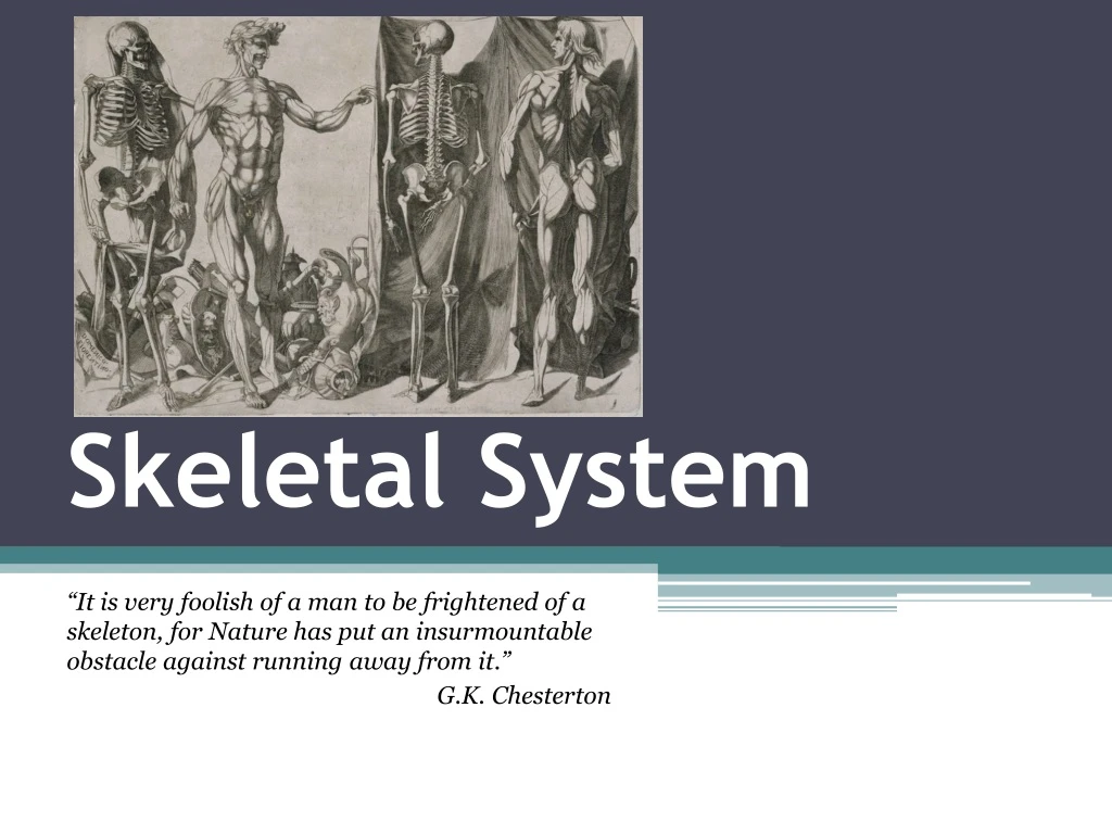
Skeletal System. “It is very foolish of a man to be frightened of a skeleton, for Nature has put an insurmountable obstacle against running away from it.” G.K. Chesterton. Functions of the Skeletal System.

E N D
Skeletal System “It is very foolish of a man to be frightened of a skeleton, for Nature has put an insurmountable obstacle against running away from it.” G.K. Chesterton
Functions of the Skeletal System • Supports the body by providing a framework for the attachment of other tissues and organs. • Storage of calcium and phosphate ions. • Production of red blood cells (in the marrow). • Protection of soft tissues and organs. • Allow movement by providing a place for muscles to attach.
Classification of Bones • Long bones are longer than they are wide, with heads at each end. • Examples include the femur and humerus.
Short bones are often cube-shaped, and contain higher amounts of spongy bone. • Examples include the bones of the wrist (carpals) and ankle (tarsals).
Flat bones are thinner, flattened, and often curved. • Made of thin layers of compact and spongy bone. • Examples include the skull, ribs, and sternum.
Irregular bonesdo not fit into any of the other categories due to their unusual shapes. • Examples include the vertebrae, sacrum, and pelvic bones.
Bone Anatomy • Bone is made of two types of tissue. • Compact bone is relatively solid, while spongy bone has many spaces in between the bony rods or struts.
Anatomy of a Long Bone • Long bones are divided into three sections: • Proximal epiphysis is the end of the bone closest (“approximate”) to the trunk of the body. • Diaphysisis the middle shaft of the bone. • Distal epiphysis is the end of the bone farthest (“distant”) to the trunk of the body.
The diaphysis is covered by a layer of dense fibrous tissue called the periosteum. • The medullary cavity is the hollowed out area inside the shaft. • Contains yellow marrow (fat storage) in adults. • Red marrow (blood cell formation) in infants.
Anatomy of a Long Bone Proximal Epiphysis Spongy Bone Compact Bone Diaphysis Medullary Cavity Periosteum Distal Epiphysis
Microscopic Anatomy of Bone • Each functional unit of bone is called an osteon. • Thin, calcified plates form the ring-like lamellae. • Each layer of a lamella contain pits called a lacunae, which contains osteocytes (bone cells).
Microscopic Anatomy of Bone • Central canals at the center of each osteon contains theblood vessels to nourish the bone. • Perforating canals join the central canals together.
Microscopic Anatomy of Bone • There are two types of bone cells. • Osteoblasts lay down the minerals needed to build the bone. • Osteoclasts shape the bone into the appropriate form. • The cells are all connected back to the nutrient supply through tiny canals called canaliculi.
Microscopic Bone Anatomy Lamella Central Canal Lamella Lacuna Osteocyte Lacuna Canaliculus
Bone Growth • Bones continuously grow and lengthen throughout childhood, up to about age 25. • In the embryonic stages of development, bones contain much more hyaline cartilage. • This cartilage is gradually replaced by bone in a process called ossification.
Hyalinecartilage New center ofbone growth Medullarycavity Bone startingto replacecartilage Bone startingto replacecartilage Bloodvessels Growthin bonelength Bone collar Bone collar Hyalinecartilagemodel Hyalinecartilagemodel In an embryo In an embryo In a fetus (a) (a) • Epiphyseal (growth) plates are layers of cartilage that are replaced by bone and regrow until adulthood. They are also called epiphyseal plates.
Articularcartilage Hyalinecartilage Spongybone New center ofbone growth New boneforming Epiphysealplatecartilage Growthin bonewidth Medullarycavity Bone startingto replacecartilage Bloodvessels Growthin bonelength New boneforming Bone collar Hyalinecartilagemodel Epiphysealplate cartilage In an embryo In a fetus In a child (a) • During adolescence, the bones grow much more quickly, overtaking the cartilage until only articular cartilage remains.
The Skeleton • The skeleton is divided into two regions: • The axial skeletonincludes everything around the longitudinal (vertical) center plane of the body. • Skull, spine • 80 bones • The appendicular skeletonincludes the appendages: the arms and legs. • Bones of arms and legs • 126 bones
The Skull • Most of the bones of the skull are flat, designed to be protective. • Each bone is joined by a suture, a joint made of dense fibrous tissue.
Fontanels • The fetal skull has a few sutures that are much wider, called fontanels. • These allow the brain to grow and expand. • Convert to bone within about 2 years. • Fontanels are the soft spots on the heads of infants.
Skull, Lateral View Parietal Bone Sphenoid Bone Frontal Bone Nasal Bone Occipital Bone Zygomatic Bone Temporal Bone Maxilla Mandible
Skull, Inferior View Maxilla Zygomatic Bone Sphenoid Bone Temporal Bone Mastoid Process Parietal Bone Occipital Bone Foramen Magnum Occipital Condyle
Sinuses • Sinusesare hollow bones with thin plates between them designed to drain fluids. • Sinus headaches happen when they get blocked and the fluids overflow into the nasal cavity.
The Hyoid Bone • The only bone in the entire body that does not form a joint with any other bone. • The base of the tongue attaches to this bone, and it aids in swallowing and speech.
Ear • The middle ear is made up of three small bones. • Malleus (hammer) • Incus (anvil) • Stapes (stirrup)
Vertebral Column • There are 24 vertebral bones, each separated by a disk of fibrocartilage. • The vertebrae are named based on their location. • C1-C7 – Cervical vertebrae in the neck • C1 is called the atlas • C2 is called the axis • T1-T12 – Thoracic vertebrae in upper back. • L1-L5 – Lumbar vertebrae in the lower back. • Two bones found below the lumbar region, made from nine vertebrae fused together. • Sacrum • Coccyx
Vertebral Column AtlasAxis CervicalC1-C7 ThoracicT1-T12 LumbarL1-L5 Sacrum Coccyx
Cervical Vertebrae Vertebral Body Vertebral Foramen Spinous Process
Thoracic Vertebrae Vertebral Body Vertebral Foramen Transverse Process Spinous Process
Lumbar Vertebrae Vertebral Body Transverse Process Vertebral Foramen Spinous Process
Ribs and Sternum • Protect major organs of the thoracic cavity. • Heart • Lungs • There are three sets of ribs: • True ribs (pairs 1-7) are connected directly to the sternum. • False ribs(pairs 8-12) are connected to the sternum through cartilage or not at all. • Floating ribs (pairs 11 and 12) are false ribs only connected to the thoracic vertebrae.
Sternum and Ribs Manubrium True Ribs: 1-7 Body of Sternum Xiphoid Process False Ribs: 8-10 Floating Ribs: 11-12
The Appendicular Skeleton Frontal Bone Parietal Bone Occipital Bone Maxilla Mandible Clavicle Scapula Sternum Humerus Rib Cage Vertebral Column Radius Sacrum Coccyx Ulna Carpals Metacarpals Phalanges
The Appendicular Skeleton Pelvis Femur Patella Tibia Fibula Talus Tarsals Metatarsals Phalanges
Scapula, Posterior View Coracoid Process Acromion Glenoid Cavity Body
Clavicle, Anterior/Superior View Sternal End Acromial End
Humerus, Right, Posterior view Head Deltoid Tuberosity Medial Epicondyle Capitulum Trochlea
Radius and Ulna Trochlear Notch Head of Radius Ulna Radius Interosseous Membrane
Right Wrist and Hand Distal Phalanges Middle Phalanges Proximal Phalanges III II IV Metacarpals I V Carpals Radius Ulna
Pelvis, Anterior View Sacrum Ilium Coccyx Pubis Acetabulum Ischium
Right Femur,Anterior Surface Neck Head Lateral Condyle Medial Condyle Patellar Surface
Right Tibia and Fibula,Anterior View Lateral Tibial Condyle Medial Tibial Condyle Head of Fibula Interosseous Membrane
Right Foot Calcaneus Tarsals V Metatarsals I Proximal Phalanges Middle Phalanges Distal Phalanges
Articulations • Articulations, also called joints, exist wherever two bones meet. • Joints are classified according to the range of motionthey allow. • Synarthrosis are immovable joints. • Amphiarthrosis are slightly moveable. • Diarthrosis are freely moveable.
Synarthroses • Immoveable joints include sutures; bones that are interlocked and bound together with dense connective tissue. • The cartilaginous connections between ribs and sternum are also immoveable.
Amphiarthroses • Bones in an amphiarthrosis are usually farther apart than a synarthrosis and can move slightly. • The interosseous membrane connecting the tibia/fibula and radius/ulna. • The symphysis pubis and the fibrocartilage between the vertebrae.