Download
1 / 53
530 likes | 1.07k Views
Learn 11 key facts about the heart, including its structure, function, location, coverings, layers, and internal anatomy. Explore the cardiac cycle, valves, sound production, blood supply, and conduction system in this comprehensive guide.

E N D
11 facts about the heart • https://www.youtube.com/watch?v=yMorctYmNUs
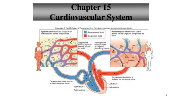
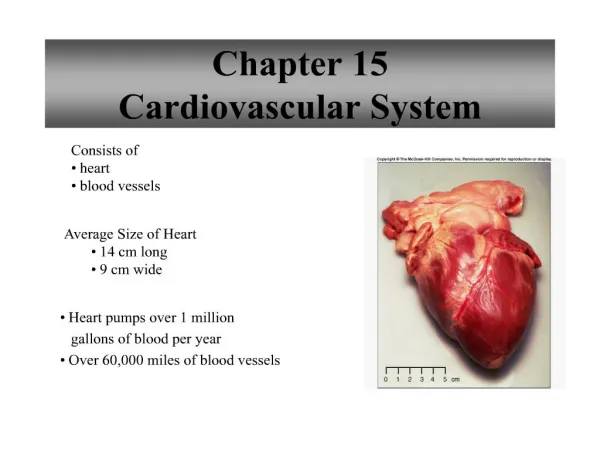
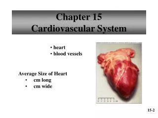
Cardiovascular System • Provides O2 & nutrients to all tissues • Removes CO2 & wastes • Consists of heart & b.v. • The heart is a 4 chambered, hollow, muscular organ • Avg. size: 9 cm wide, 14 cm long
Location • Located in the mediastinum, resting on the diaphragm • Base – top of heart beneath 2nd rib • Apex – point at bottom (points toward left) at 5th intercostal space • Bordered by lungs laterally & sternum anteriorly
Coverings • Fibrous pericardium – tough, outer layer • Parietal pericardium – inner lining of pericardial cavity Pericardial cavity – space filled w/serous fluid; reduces friction 3. Visceral pericardium – membrane that covers the heart (same as epicardium)
Coverings Fibrous pericardium→ Fibrous Visceral pericardium pericardium
Layers of Heart Wall 1. Epicardium – same as vis. pericardium; thin connective tissue; protection & secretes fluid 2. Myocardium – thick; consists of cardiac muscle; contracts to pump blood 3. Endocardium – epithelial & connective tissue; lines all chambers & valves; helps keep surfaces smooth
Heart – External Features • Auricle – ext. flaps • Sulci – grooves for b.v. • During an avg. lifetime, the heart beats approx. 2.5 billion times • It pumps an avg. of approx. 300 million L of blood
Heart Animation • Heart Animation • Heart Crash Course
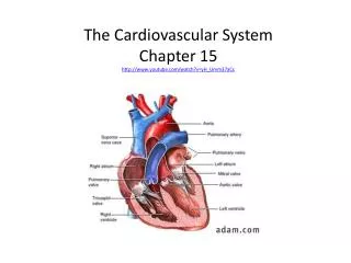
Internal Anatomy • The heart has been described as 2 pumps that lie side by side • The 2 sides of the heart are separated by a wall called the septum • The 2 thinner-walled upper chambers that receive blood are called atria • The 2 thicker-walled bottom chambers that pump blood are called ventricles
Internal Anatomy • The atria are separated from the ventricles by valves • Valves prevent the backflow of blood • Tricuspid – on rt. • Bicuspid (mitral) on left • Rt. side of heart receives deoxy. blood from body & pumps it to the lungs to get oxygenated (colored blue) • Left side receives oxygenated blood from lungs & pumps it to body (colored red)
Heart Valves • 2 types: 1. Atrioventricular valves (A-V valves) – located between the 2 atria & ventricles: tricuspid & bicuspid (mitral) 2. Semilunar valves (S-L valves) – located in the b.v. leading away from the heart: pulmonary & aortic
Chordae Tendinae • In the right and left ventricle chordae tendinae are strong fibers attached to the A-V valves. • They prevent the valve from extending into the atrium chamber. • Attached to papillary muscles.
Heart Sounds • Produced by the closing of the heart valves • A typical heartbeat is heard as “lub-dup” • A-V sounds: • Mitral – heard at 5thintercostal space • Tricuspid – at tip of sternum • S-L sounds: • Aortic – at 2nd intercostal space on rt. • Pulmonary – 2nd intercostal space on left
Aortic Valve Area Second right intercostal space (ICS), right sternal border • Pulmonic Valve Area Second left intercostal space (ICS), left sternal border • Tricuspid Valve Area Fourth left ICS, left sternal border • Mitral Valve Area Fifth ICS, left mid-clavicular line
Blood Supply to the Heart (anterior view) • The left & rt. coronary arteries branch off the aorta to supply blood to the heart itself • The l.c. art. branches into circumflex & left ant. descending artery (or left interventricular artery)
Blood Supply to the Heart(anterior view) • The rt. coronary artery branches into the marginal artery & the posterior inter- ventricular artery (on posterior view – next slide)
Blood Supply to the Heart • Blood leaves arteries & passes through capillaries of the myocardium • Blood then passes into cardiac veins • Cardiac veins empty into coronary sinus (lg. vein on post. side of heart) • Coronary sinus empties into rt. atrium
Cardiac Conduction System • The heart has its own pacemaker that can initiate a heart- beat; called the sinoatrial node (S-A node) • Located in the upper rt.atrium; causes both atria to contract almost simultaneously
Cardiac Conduction System • Impulse travels to the atrioventricular node (A-V) located in the interatrial septum • Impulse then moves to A-V bundle (or bundle of His) & then to bundle branches • Purkinje fibers carry impulseto distant parts of ventricles
Cardiac Conduction System • Cardiac Conduction System
The Cardiac Cycle • A series of contractions & relaxations that constitute one heartbeat • Atria contract while ventricles relax & vice versa • Diastole – relaxation of a heart chamber • Systole – contraction of a heart chamber
Cardiac Cycle Animation • Cardiac Cycle Animation
Electrocardiograms (EKGs) • Measures electrical activity of the heart • Results from depolarization & repolarization of the myocardium • 12 electrodes are placed on various places on the body • Changes can indicate arrhythmias & other heart-related conditions
Parts of an EKG • P wave – contraction of the atria • QRS complex – contrac- tion of ventricles • T wave – relaxation of ventricles • Relaxation of atria (repolarization) – not seen (hidden by QRS complex)
EKG Animation • EKG Animation
EKG Measurements • P wave to P wave - 1 complete ♥ beat • P-Q interval – time it takes impulse to travel from S-A node to A-V node
EKG Measurements • Q-T interval - the period from the beginning of ventricular depolarization until the end of ventricular repolarization • The QT interval is prolonged if it clearly measures more than half the R-R interval • Called long QT syndrome
Abnormal EKGs • Long QT syndrome
Bundle Branch Block • The QRS complex is the time required to depolarize the ventricles. • A normal QRS is 0.08-0.12 s • > than 0.12 seconds is considered a BBB (block in a bundle branche, or the electrical impulse was conducted abnormally)
Arrhythmias • Arrhythmias
defibrillator • Defibrillator video
Arrhythmias • Normally the ♥ contracts about 60-100 times/min • Each contraction represents one ♥ beat • Abnormal ♥ rates & EKGs – called arrhythmias
Arrhythmias Sinus rhythm → 60-80 beats/min Tachycardia > 100 beats/min Bradycardia < 60 beats/min
Arrhythmias • Atrial Flutter – many more P waves compared to QRS complexes • Atrial Fibrillation – P waves absent
Arrhythmias • Junctional Rhythm – S-A node nonfunctional; P waves absent; heart being paced by A-V node (40-60 beats/min) • Ventricular Fibrillation – Chaotic depolarization of ventricles; extremely irreg. as seen in heart attack, elec. shock
Arrythmias • Asystole- no heart activity
Blood Pressure • Pulse – alternate expansion & relaxation of arteries • Pulse pressure – difference b/t systolic & diastolic pressure • Central venous pressure – pressure in rt. atrium
Factors That Affect B.P.BP An incr. in any of these causes an incr. in BP: 1.Heart Action • Cardiac Output (CO) = Vol. of blood discharged from the ventricles/min. • Stroke Volume (SV) = Vol. of blood discharged w/each contraction (approx. 70 ml at rest) • Heart Rate (HR) = number of beats/min • CO = SV X HR • CO = 70 ml X 70 beats/min • CO = 4900 ml/min
Factors That Affect B.P.P • Blood Volume = sum of formed elements + plasma (usually 5L); (ex. - hemorrhage results in drop in B.P.) 3. Peripheral Resistance – force produced from the friction on blood from b.v. walls (expansion or dilation of b.v. changes resistance) • Viscosity = ease w/which blood flows (an incr. of RBCs incr. viscosity; anemia or hemorrhage decr. it)
Blood Pressure • Force blood exerts against inner walls of b.v. (usually arteries) • Avg. = 120/80 • Measured w/a sphygmomanometer • 120 = systolic – maximum pressure during ventr. contraction • 80 = diastolic – lowest pressure during ventr. relaxation
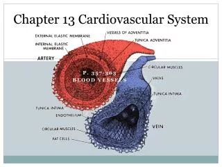
Blood Vessels • Include arteries, veins & capillaries • Arteries & veins have 3 layers: 1. tunica intima – single layer squamous cells; secretes substances to stem blood flow 2. tunica media – makes up most of the b.v.; mostly smooth muscle & some connective tissue; allows for expansion 3. tunica externa – connective tissue; provides attachment to surrounding tissue
Blood Vessels • 2 differences b/t arteries & veins: 1. thickness of tunica media 2. veins have valves
Capillaries • Smallest b.v.; connect arterioles to venules • Resp. gases & other substances are exchanged in capillaries thru diffusion
Movement In Capillaries • Water & other substances leave capillaries at the arteriolar end b/c of a net outward pressure • Water & other substances enter at venular end b/c of a net inward pressure