Chapter 20: The Cardiovascular System
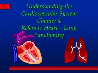
Chapter 20: The Cardiovascular System. THE HEART. Heart Anatomy. Location diaphragm, mediastinum, 2/3 left of midline Orientation Apex- points anterior, inferior, left Base- directed posterior, superior, right Vessels Superior and Inferior Vena Cava

Chapter 20: The Cardiovascular System
E N D
Presentation Transcript
Chapter 20: The Cardiovascular System THE HEART
Heart Anatomy • Location • diaphragm, mediastinum, 2/3 left of midline • Orientation • Apex- points anterior, inferior, left • Base- directed posterior, superior, right • Vessels • Superior and Inferior Vena Cava • Pulmonary trunk pulmonary arteries(lungs) • Pulmonary veins • Aorta
Pericardium- figure 20.2 • Membrane that surrounds & protects • Confines to position in mediastinum • 2 main parts: • Fibrous pericardium- superficial, anchor • Tough, inelastic, dense irregular CT • Baglike, open end attached to vessels • Prevents overstretching of heart • Serous pericardium- thinner, delicate • Forms double layer (pericardial fluid in pericardial cavity - reduces friction, allows movement): • Parietal layer- fused to fibrous • Visceral layer- inner = EPICARDIUM- adheres tightly to heart surface
Layers of the heart wall • Epicardium- thin, transparent, outer • Visceral layer of serous pericardium • Smooth slippery outside of heart • Myocardium- middle • Cardiac muscle- striated but involuntary • Bulk of heart • Pumping action • Endocardium- inner • Thin endothelium over CT • Smooth lining of chambers and valves • Continuous with b.v.
Heart Anatomy fig 20.3-6 • Heart chambers = 4 • 2 Atria • Right- receives blood from vena cavae • Left- receives blood from pulmonary veins • 2 Ventricles • Right- pumps deoxygenated blood to lungs • Left- pumps oxygenated blood to systemic circ • Myocardium much thicker than right ventricle • Heart valves = 4 • Atrioventricular valves = tricuspid & bicuspid • Semilunar valves = aortic and pulmonary
Valve function • When AV valve open: • Cusps project into ventricle • Ventricle relaxed papillary muscle relaxed chordae tendineae slack • Blood: pressure atria pressure ventricle • Ventricle contracts, pressure cusps up, close • Papillary muscles contract chordae tendineae tighten • SL valves openwhen pressure in ventricles exceeds pressure in arteries • As ventricles relax blood moves back toward heart SL valves close
Terms • Auricles – on anterior surface of atria • Increases capacity of each atrium so each can hold a greater volume of blood • Coronary sulcus – separation between atria and ventricles • Systole – contraction • Diastole – relaxation • Tachycardia – high heart rate, > 100bpm • Bradycardia – low heart rate, 50 bpm
Coronary circulation (2) • Coronary – “crown,” encircles heart • contracts, little blood flows coronary artery but as relaxes, aorta pushes blood thru coronary arteries • Anastomoses – area where 2 or more arteries supply the same region • Provide alternate routes for blood to reach a particular organ or tissue • Myocardium contains • Provides detours if main route is obstructed
Problems… • Myocardial ischemia – partial obstruction of blood flow in coronary arteries • blood flow to myocardium • hypoxia may weaken cells w/out killing them • Silent = episodes without pain, dangerous in that no forewarning to attack • Angina pectoris – “strangled chest” • Severe pain usually accompanies myocardial ischemia • Tightness or squeezing sensation • Can occur during exertion when requires more O2 • Pain referred to neck, chin, left arm
Myocardial infarction (MI) • Heart attack • Complete obstruction of blood flow to coronary artery • Infarction = death of tissue area due to interrupted blood supply • Tissue distal to obstruction dies, replaced by non-contractile scar tissue loses strength • May also disrupt conduction system and cause sudden death – ventricular fibrillation – rapid uncoordinated twitching that disrupts regular rhythm • treatment: injection of clot dissolver, plus heparin, coronary angioplasty or coronary artery bypass
Properties of cardiac muscle cells • Shorter than skeletal • Branching • Central nucleus, sometimes binucleate • Intercalated discs- thickenings of sarcolemma, contain: • Desmosomes- hold fibers together • Gap junctions- for AP conduction • Mitochondria large & numerous • Like skeletal- arrangement of proteins • SR smaller less intracellular Ca2+ • T-tubules wider but less abundant
Functional syncytium • stimulation of individual muscle cell results in contraction of all muscle cells due to gap junctions in intercalated discs • an application of the all-or-none principle • If stimulus in cardiac muscle is great enough to initiate contraction of a single cell, the entire muscular syncytium will undergo contraction
Contraction physiology • 1% of cardiac fibers become autorhythmic during embryonic development • Pacemaker function- set rhythm of electrical excitation • Conduction system- network of specialized fibers provide path for excitation to progress thru heart • Ensuring coordinated contraction of chambers • Both atria contract at same time • Both ventricles contract at same time • Cardiac AP goes thru following sequence…
Contraction physiology (2) • Pathway of stimulation • 1. Sinoatrial (SA) node- cells do not have a stable resting membrane potential • depolarized spontaneously = pacemaker potential • 2. Atrioventricular (AV) node • 3. Bundle of His • 4. Bundle branches • 5. Purkinje fibers • 6. Ventricular cells- contraction pushes blood up to SL valves
Cardiac Action Potentials, 20.11 • Depolarization: Na+ gates open= fast channels • Rapid depolarization because they open fast • Plateau: opening of slow Ca2+ channels in the sarcolemma • More Ca2+ outside cell cytosol also causing Ca2+ to pour out of SR • Ca2+ contraction • K+ channels opening but Ca2+ balances it remains depolarized for about 0.25 sec • (in skeletal muscle 0.001 sec, no plateau phase) • Repolarization: K+ outflow restores resting m.p. • Ca2+ channels also are closing
Cardiac Action Potentials (2) • Positive inotropic agents contractility (substances promote inflow of Ca2+ channels strength contractions • NE and Epinephrine modify • Timing • strength of contraction • Do NOT establish a rhythm • Digitalis • interstitial Ca2+ • Negative inotropic agents contractility • Ach released by Parasymp NS slows SA node pacing from 100 to about 75 AP/minute • Also: anoxia, acidosis, some anesthetics, K+, Ca2+ channel blockers
Long refractory pd- cardiac muscle • Refractory pd- time interval during which second contraction cannot be triggered • In cardiac- longer than contraction pd • Another contraction cannot happen until relaxation is happening • Tetanus cannot occur • If tetanus occurred blood flow would cease
Arrhythmias • Irregular rhythm due to conduction defect • Causes: • Caffeine, nicotine, alcohol, other drugs, anxiety, hyperthyroidism, K+ deficiency, & some heart disease • Examples: • Heart block – AP slowed or blocked (3 types) • 1st °= AP slow thru AV, 2nd °= some AP not thru AV node, 3rd ° = no AP thru AV node • Atrial flutter – rapid atrial contractions • Atrial fibrillation – asynchronous cont- atrial fibers • Ventricular fibrillation– async cont ventricular fibers* • Premature ventricular contraction – ectopic area of high excitation abnormal AP (before SA node intends)
Electrocardiogram (ECG) • P wave – atrial depolarization atrial contraction ventricular filling • QRS complex – ventricular depolarization ventricular contraction SL valves open blood ejection • Rt ventriclepulmonary trunk pul arteries lungs • Left ventricle aorta systemic circulation • T wave – ventricular repolarization