The Cardiovascular System CHAPTER 8
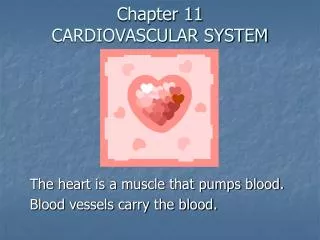
The Cardiovascular System CHAPTER 8. Pump it Up!!!. The heart is a pump that moves blood through the body. This movement allows oxygen, nutrients, hormones, antibodies , and inflammatory cells to be delivered to where they are needed in the body.

The Cardiovascular System CHAPTER 8
E N D
Presentation Transcript
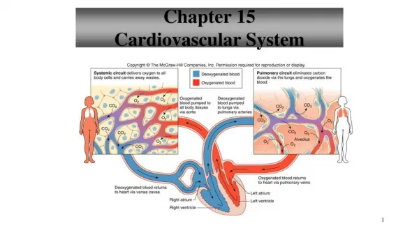
Pump it Up!!! • The heart is a pump that moves blood through the body. This movement allows oxygen, nutrients, hormones, antibodies, and inflammatory cells to be delivered to where they are needed in the body. • Also removes__________ products from tissues. • All of these materials are propelled by the heart through a closed system of tubes to the tissues of the body.
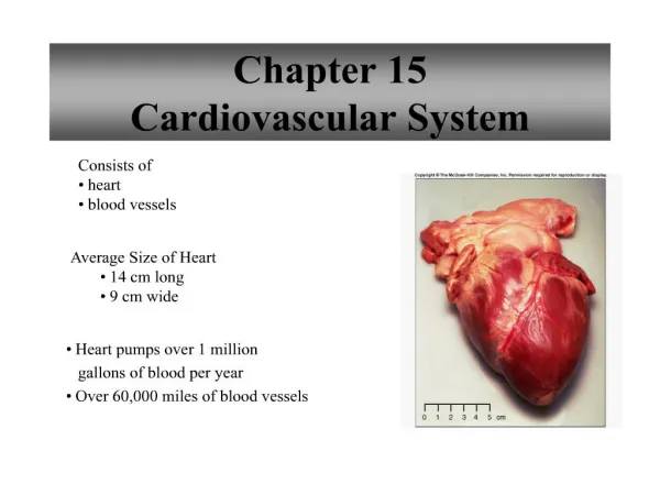
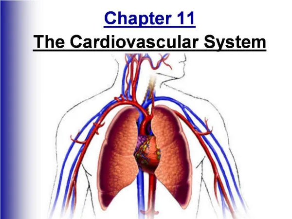
Where is the heart located? • Centrallyin the chest • Surrounded by lungs, protected by ribs • Between 3rd-7thribs in ________ animals and 2nd-6thribs in _________ animals • Lies in the _____________/interpleural space, which is the space between the pleural covering of the right and left lungs. • Trachea, esophagus, and other vascular structures are also contained in the mediastinum.
Orientation of the heart • Shifted to the left of the chest • ________ - the bottom of the heart where the left ventricle comes to a point • ________ - the top of the heart where major blood vessels enter and exit
Composition of the Heart Wall Outer layer of the heart is called the_________________. The pericardium consists of an outer fibrous pericardium and an inner serous pericardium. • ______________ pericardium • Made of tough, fibrous connective tissue that protects the heart and loosely attaches to the diaphragm.
The ____________ pericardium consists of two layers: • *The __________ pericardial layer is adjacent to the fibrous pericardium. • *The __________ pericardial layer (epicardium) is adhered to the myocardium.
Between the two layers of the serous pericardium is the pericardial space. This is a fluid filled cavity that ________________ the two layers which allows the heart to smoothly expand and contract.
Beneath the epicardium is the ______cardium, the thickest layer of the heart tissue. • Between the myocardium and the heart chambers is a thin membranous lining called the ______cardium.
External anatomy of the heart: Chambers • Auricles- (left and right) largest and most visible parts of the ________. They are identified by knowing which ventricle they lie above. • ________ ventricle - long and narrow, thick-walled, terminates at apex of heart • ________ ventricle - broader surface area, wraps around left ventricle • The borders of the ventricles are separated by interventricular sulci, which are grooves that contain fat and blood vessels that are part of ____________ circulation of heart.
External anatomy of the heart: Vessels carrying DEOXYGENATED blood • The _______ _________ (cranial and caudal) empty deoxygenated blood into the right atrium • The Pulmonary _________ emerges from the right ventricle as the pulmonary trunk • Quickly divides into right and left pulmonary arteries traveling to each lung
External anatomy of the heart: Vessels carrying OXYGENATED blood • The Pulmonary _________ (left and right) return oxygenated blood to the left atrium • The _________ is the largest artery in the body • The walls of the aorta are the thickest of any blood vessel • The aorta emerges from the left ventricle into the aortic arch
Internal Structures of the Heart Atrioventricular Valves: -Right Atrioventricular Valve (aka Right AV valve/Tricuspid Valve) -has ___ flaps/leaflets -Left Atrioventricular Valve (aka Left AV Valve/Mitral Valve/Bicuspid Valve) -has ___ flaps/leaflets The AV valves are attached to _________ __________, which prevent the flaps from bending back into the atria. Chordae tendinae connect the free edges of the valvular flaps to the papillary muscles. _____________ muscles attach to the interventricular septum
The _____________ band is a band of tissue present in the right ventricle. -originates at interventricular septum -not attached to flaps of tricuspid valve -provides additional structural support to the wall of the right ventricle
Semilunar valves: -Aortic valve -Pulmonary/pulmonic valve Both valves are made of ____ leaflets. The aortic valve leads to the aorta and the pulmonary valve leads to the pulmonary artery.
Closure of the AV valves prevent blood from flowing backward into the atria. Closure of the semilunar valves prevent blood from flowing backward into the.The flaps of all 4 valves originate from a fibrous ring of tissue called the ___________.