Download
1 / 45
540 likes | 867 Views
PBIO 691: Seminar (Plant Cell Walls) . Dr. Allan Showalter Department of Environmental & Plant Biology Molecular and Cellular Biology Program Ohio University Athens, OH 45709. Introduction. There are three major regions of the wall:

E N D
PBIO 691: Seminar(Plant Cell Walls) Dr. Allan Showalter Department of Environmental & Plant Biology Molecular and Cellular Biology Program Ohio University Athens, OH 45709
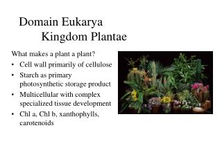
Introduction There are three major regions of the wall: • Middle lamella - outermost layer, glue that binds adjacent cells, composed primarily of pectic polysaccharides. • Primary wall - wall deposited by cells before and during active growth. The primary wall of cultured sycamore cells is comprised of pectic polysaccharides (ca. 30%), cross-linking glycans (hemicellulose; ca 25%), cellulose (15-30%) and protein (ca. 20%) (see Darvill et al, 1980). The actual content of the wall components varies with species and age. All plant cells have a middle lamella and primary wall. • Secondary Wall - some cells deposit additional layers inside the primary wall. This occurs after growth stops or when the cells begins to differentiate (specialize). The secondary wall is mainly for support and is comprised primarily of cellulose and lignin. Often can distinguish distinct layers, S1, S2 and S3 - which differ in the orientation, or direction, of the cellulose microfibrils.
The Plant Cell Wall a | Cell wall containing cellulose microfibrils, hemicellulose, pectin, lignin and soluble proteins.b | Cellulose synthase enzymes are in rosette complexes, which float in the plasma membrane.c | Lignification occurs in the S1, S2 and S3 layers of the cell wall.
Functions of the plant cell wall • maintaining/determining cell shape • support and mechanical strength • prevents the cell membrane from bursting in a hypotonic medium • controls the rate and direction of cell growth and regulates cell volume • ultimately responsible for the plant architectural design and controlling plant morphogenesis • has a metabolic role • physical barrier to: (a) pathogens; and (b) water in suberized cells. However, remember that the wall is very porous and allows the free passage of small molecules, including proteins up to 60,000 MW. • carbohydrate storage - the components of the wall can be reused in other metabolic processes (especially in seeds) • signaling - fragments of wall, called oligosaccharins, act as hormones • recognition responses - for example: (a) the wall of roots of legumes is important in the nitrogen-fixing bacteria colonizing the root to form nodules; and (b) pollen-style interactions are mediated by wall chemistry • economic products - cell walls are important for products such as paper, wood, fiber, energy, shelter, and even roughage in our diet
2.1 Sugars: building blocks of the cell wallThe monosaccharides in cell wall polymers are derived from glucose.
2.2 Macromolecules of the cell wall • Cellulose • Callose • Hemicellulose • Pectin • Cell wall protein • Phenolics
2.2.1 Cellulose is the principal scaffoldingcomponent of all plant cell walls. • Cellulose in primary and secondary cell walls • Made of (1→4)β-D-glucan chains hydrogen bonded to one another along their length • Groups of 30 to 40 of these chains laterally hydrogen bond to form crystalline or para-crystalline microfibrils
Callose • Callose differs from cellulose in consisting of (1→3)β-D-glucan chains, which can form helical duplexes and triplexes • Callose is made by a few cell types at specific stages of wall development, such as in growing pollen tubes and in the cell plates of dividing cells • Callose is also made in response to wounding
Hemicellulose • Cross-linking glycans interlock the cellulosic scaffold • Cross-linking glycans are a class of polysaccharides that can hydrogen-bond to cellulose microfibrils and link them together to form a network • Most cross-linking glycansare often called “hemicelluloses,” a widely used but archaic term for all materials extracted from the cell wall with molar concentrations of alkali, regardless of structure • Includes Xyloglucans (XyGs), Xylans [glucuronoarabinoxylans (GAXs), arabinoxylans (AXs), glucuronoxylans(GXs)], Mannans (glucomannans, galactoglucomannans, and galactomannans), and “mixed-linkage” (1→3),(1→4)β-D-glucans (β-glucans)
Xyloglucans and glucuronoarabinoxylans • The two major cross-linking glycans of all primary cell walls of flowering plants are xyloglucans (XyGs) and glucuronoarabinoxylans(GAXs) (Fig. 2.12). • XyGs crosslink the walls of all dicots and about one halfof the monocots, but in the cell walls of the “commelinoid” line of monocots, which includes bromeliads, palms, gingers, cypresses, and grasses, the major cross-linking glycan is GAX (Fig. 2.13).
In the order Poales, which contains the cereals and grasses, a third major crosslinkingglycan, called “mixed-linkage” (1→3),(1→4)β-D-glucans (β-glucans), distinguishesthese species from the other commelinoid species (Fig. 2.14).
2.2.3 Pectin matrix polymers are rich in galacturonic acid • Pectins—a mixture of heterogeneous, branched, and highly hydrated polysaccharides rich in D-galacturonic acid—have been defined classically as material extracted from the cell wall by Ca2+-chelatorssuch as ammonium oxalate, EDTA, EGTA, or cyclohexanediaminetetraacetate. • Two fundamental constituents of pectinsare homogalacturonan (HGA; Fig. 2.16A) and rhamnogalacturonan I (RG I; Fig. 2.16C). • There are two kinds of structurally modified HGAs, xylogalacturonan(Fig. 2.16B) and rhamnogalacturonanII (RG II; Fig. 2.17A). • Other polysaccharides, composed mostly of neutral sugars—such as arabinans, galactans, and highly branched type I arabinogalactans(AGs) of various configurations and sizes—are attached to the O-4 of many of the Rha residues of RG I (see Fig. 2.16D).
2.2.4 Structural proteins of the cell wall are encoded by large multigene families (Figs. 2.18 and 2.20) • The hydroxyproline-rich glycoproteins (HRGPs) include: • Extensins (EXT) • Proline-rich proteins (PRPs) • Arabinogalactan-proteins (AGPs) • In addition, there are the glycine-rich proteins (GRPs)
2.2.5 Aromatic substances are present in the nonlignified walls of commelinoid species • A large fraction of plant aromatics consists of hydroxycinnamic acids, such as ferulic and p-coumaric acids (Fig. 2.21; see also Chapter 24). • In grasses, these hydroxycinnamatesare attached as carboxyl esters to the O-5 position of a few of the Ara units of GAX. A small proportion of the ferulic acid units of neighboring GAXs may cross-link by phenyl–phenyl or phenyl–ether linkages to interconnect the GAX into a large network (Fig. 2.22).
2.3 Cell wall architecture • The primary wall consists of three structural networks: • Cellulose and cross-linking glycans • Matrix pecticpolysaccharides • Structural proteins or a phenylpropanoid network
2.3.2 Walls of angiosperms are arranged in two distinct types of architecture • Type I walls- most dicots and the noncommelinoid monocots contain about equal amounts of XyGs and cellulose • In Type I walls (Fig. 2.23A), the cellulose-XyG framework is embedded in a pectin matrix that controls, among other physiological properties, wall porosity. HGA is thought to be secreted as highly methyl-esterified polymers, and the enzyme pectin methylesterase(PME), located in the cell wall, cleaves some of the methyl groups to initiate binding of the carboxylateions to Ca2+. • Type II walls- commelinoid monocots contain cellulosemicrofibrils similar to those of the Type I wall; instead of XyG, however, the principal polymers that interlock the microfibrilsare GAXs. • In general, Type II walls are pectin-poor, but an additional contribution to the charge density of the wall is provided by the α-L-GlcA units on GAX. These walls have very little structural protein compared with dicots and other monocots but they can accumulate extensive interconnecting networks of phenylpropanoids, particularly as the cells stop expanding.
2.4 Cell wall biosynthesis and assembly • Cell walls originate in the developing cell plate. As plant nuclei complete division during telophase of the mitotic cell cycle, the phragmosome, a flattened membranous vesicle containing cell wall components, forms across the cell within a cytoskeletal array called the phragmoplast. • Cell wall synthesis continues after cell division. • The plant Golgi apparatus is a factory for the synthesis, processing, and targeting of glycoproteins (Fig. 2.26). • The Golgi apparatus also has been shown by autoradiography to be the site of synthesis of noncellulosic polysaccharides. • Thus—except for cellulose—the polysaccharides, the structural proteins, and a broad spectrum of enzymes are coordinately secreted in Golgi-derived vesicles and targeted to the cell wall.
2.4.2 Golgi-localized enzymes interconvert the nucleotide sugars, which serve as substrates for polysaccharide synthesis • The reactions that synthesize noncellulosiccell wall polysaccharides in the Golgi apparatus utilize several nucleotide sugars as substrates. • Beginning with formation of UDP-glucose and GDP-glucose (Fig. 2.27), pathways for nucleotide sugar interconversionproduce various nucleotide sugars de novo in enzyme-catalyzed reactions (Figs. 2.28 and 2.29). • Many of these interconversionenzymes (e.g., epimerases and dehydratases) appear to be membrane-bound and localized to the ER-Golgi apparatus.
2.4.4 Cellulose microfibrils are assembledat the plasma membrane surface • The only polymers known to be made at the outer plasma membrane surface of plants are cellulose and callose. • Cellulose synthesis is catalyzed by multimeric enzyme complexes located at the termini of growing cellulose microfibrils(Fig. 2.31). • These terminal complexes are visible in freeze-fracture replicas of plasma membrane. In some algae, terminal complexes are organized in linear arrays, whereas in others—and in all angiosperms—they form particle rosettes (Fig. 2.31). • Why do plants have so many different CesAand related genes?
2.5 Growth and cell walls • During elongation or expansion, existing cell wall architecture must change to incorporate new material, increasing the surface area of the cell and inducing water uptake by the protoplast. • The regulation of wall loosening is considered the primary determinant of rates of cell expansion. • The acid-growth hypothesis postulates that auxin-dependent acidification of the cell wall promotes wall extensibility and cell growth. • At present, two kinds of enzymes are being evaluated as having possible wall-loosening activities. • Xyloglucanendotransglycosylase(XET), carries out a transglycosylation of XyG in which one chain of XyG is cleaved and reattached to the nonreducing terminus of another XyG chain. • Expansins, these proteins probably catalyze breakage of hydrogen bonds between cellulose and the load-bearing cross-linking glycans.
2.6 Cell differentiation • Fruit-ripening involves developmentally regulated changes in cell wall architecture (pectin methyl esterase and polygalacturonase I and II) • Secondary walls are elaborated after the growth of the primary wall has stopped • The cotton fiber, for example, consists of nearly 98% cellulose at maturity • The secondary wall may, however, contain additional noncellulosic polysaccharides, proteins, and aromatic substances such as lignin. • Secondary deposition of suberinand cutin can render cell walls impermeable to water
Lignin biosynthetic pathway, simplified schematic. Ralph J et al. PNAS 1998;95:12803-12808 ©1998 by The National Academy of Sciences
Possible mechanisms and cellular locations of cutin and suberin assembly
2.7 Cell walls for food, feed, fuel, and fibers • Wood • Paper • Textiles • Fruits and vegetables for humans and animals • Jams, jellies, thickening agents, emulsifiers • Dietary fiber • Biomass • Biofuel: cellulosic ethanol
Cellulosic Ethanol Production and Research Challenges This figure depicts some key processing steps in a future large-scale facility for transforming cellulosic biomass (plant fibers) into biofuels. Three areas where focused biological research can lead to much lower costs and increased productivity include developing crops dedicated to biofuel production (see step 1), engineering enzymes that deconstruct cellulosic biomass (see steps 2 and 3), and engineering microbes and developing new microbial enzyme systems for industrial-scale conversion of biomass sugars into ethanol and other biofuels or bioproducts (see step 4). Biological research challenges associated with each production step are summarized in the right portion of the figure.
Model systems to study plant cell walls Switchgrass (Panicumvirgatum) Arabidopsis thaliana Brachypodiumdistachyon Miscanthusgiganteus
Methods used to study plant cell walls • Carbohydrate composition and linkage analysis • Gas chromatography-mass spectrometry (GC-MS) • Nuclear magnetic resonance (NMR) • Mass Spectrometry (MS) • Sequence-dependent glycanases • High performance liquid chromatography (HPLC) • High-pH anion-exchange chromatography (HPAC) or “Dionex” • Pulsed amperometric detector (PAD) • Microscopy (LM, SEM, TEM, AFM, FTIR, FT-Raman) • Plant tissue culture • Antibodies • Molecular biology • Molecular genetics • Biochemistry • Mutants • Transgenic plants
Resources for plant cell wall research • Carbohydrate-Active enZYmes Database (CAZy)-http://www.cazy.org/ • Glycoside Hydrolases (GHs) : hydrolysis and/or rearrangement of glycosidic bonds • GlycosylTransferases (GTs) : formation of glycosidic bonds • Polysaccharide Lyases (PLs) : non-hydrolytic cleavage of glycosidic bonds • Carbohydrate Esterases (CEs) : hydrolysis of carbohydrate esters • Antibodies for plant cell wall polymers-http://www.ccrc.uga.edu/~mao/wallmab/Antibodies/antib.htm and http://www.plantprobes.net/
References • Carpita, N. and McCann, M. (2000) Cell Walls. Chapter 2. In Biochemistry & Molecular Biology of Plants. Buchanan, BB, Gruissem W, Jones, RL. eds. American Society of Plant Biology, Beltsville, MD. • Albersheim P., Darvill A., Roberts K., Sederoff R., Staehelin A. (2011) Plant Cell Walls. Garland Science, New York.