Download

1 / 18
180 likes | 222 Views
This lecture discusses the analysis and interpretation of synovial fluid in the hematology laboratory, covering aspects such as appearance, viscosity, cell counts, crystal identification, and more. Learn about normal versus abnormal findings, the significance of viscosity, and diagnostic methods for arthritis and crystal-induced conditions.

E N D
URINALYSIS AND BODY FLUIDS (SYNOVIAL FLUID)LECTURE TWO Dr. Essam H. Jiffri
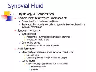
Synovial Fluid in the Haematology Laboratory • - The major portion of the routine synovial fluid analysis takes place in the hematology laboratory and includes a report of the appearance, viscosity, cell count, cell differential, and crystal identification. • - Normal synovial fluid appears clear and pale yellow.
Synovial Fluid in the Haematology Laboratory • The color becomes a deeper yellow in the presence of inflammation and may have a greenish color with bacterial infection. • Turbidity occurs when the cell count is elevated and is usually proportional of the number of cells present. • A milky fluid may also indicate the presence of crystals.
Synovial Fluid in the Haematology Laboratory • Viscosity of the synovial fluid comes from the polymerization of the hyaluronic acid and is essential for the proper lubrication of the joints. • - Arthritis affects both the production of hyaluronate and its ability to polymerize, thus decreasing the viscosity of the fluid.
Synovial Fluid in the Haematology Laboratory • Several methods are used to measure the viscosity of the fluid, the simplest being to observe the ability of the fluid to form a string from the tip of a syringe. • - A string that measures 4 to 6 cm is considered normal.
Synovial Fluid in the Haematology Laboratory • - The laboratory may be requested to measure the degree of hyaluronate polymerization by performing a Ropes, or mucin clot, test, when added to a solution of 2 to 5 percent acetic acid, normal synovial fluid will form a solid clot surrounded by clear fluid.
Synovial Fluid in the Haematology Laboratory • As the ability of the hyaluronate to polymerize decreases, the clot becomes less firm and the surrounding fluid increases in turbidity. • - The Ropes test is reported in terms of good (solid clot), fair (soft clot), poor (friable clot), and very poor (no clot).
Synovial Fluid in the Haematology Laboratory • - Both red and white blood cell counts should be performed on all specimens. • - Manual counts on the Neubauer counting chamber in the same manner as CSF and sperm counts. • - Clear fluids can usually be counted undiluted, but dilutions are necessary when fluids are turbid or bloody.
Synovial Fluid in the Haematology Laboratory • Electronic cell counters should not be used for synovial fluid cell counts because the viscous fluid may clot the tubing and the presence of tissue cells can falsely elevate counts. • - The normal synovial fluid red blood cell count is 0 to 2000 cells per microliter.
Synovial Fluid in the Haematology Laboratory • - White blood cell counts below 200 cells per microliter are considered normal and may reach 100,000 cells per microliter or higher in severe inflammations. • - Mononuclear cells, including lymphocytes, monocytes, macrophages, and synovial tissue cells, are the primary cells seen in normal synovial fluid.
Synovial Fluid in the Haematology Laboratory • - Neutrophils should account for less than 25 percent of the differential count. • Differential counts should be performed on thinly smeared Wright-stained slices.
Summary of Laboratory Findings in Joints Disorders • _______________________________________________ • Group Classification Laboratory Findings • ________________________________________________________ • I. Noninflamatory Clear, yellow fluid, good viscosity, WBCs < 5000, neutrophils < 30% • II. Inflammatory Cloudy, yellow fluid, poor viscosity, WBCs 2,000 to 100,000, neutophils > 50%, possible auto-antibodies present • Septic Cloudy, yellow-green fluid, poor viscosity, WBCs 10,000 to 200,000, • neutophils > 90%, possible culture • Crystal-induced Cloudy or milky fluid, poor viscosity, WBCs 500 to 200,000, neutophils < 90%, elevated uric acid, crystals present • V. Hemorrhagic Cloudy or red fluid, poor viscosity, • WBCs < 5000, neutrophils < 50%, • RBCs present
Summary of the most frequently encountered cells and inclusions seen in the synovial fluid
Synovial Fluid in the Haematology Laboratory • Microscopic examination of synovial fluid for the presence of crystals is used in the diagnosis of crystal-induced arthritis. • - The primary crystals seen in synovial fluid are monosodium urate (uric acid), which is found in cases of gout, and calcium pyrophosphate, which is associated with cases of pseudogout.
The microscopic characteristics of the synovial fluid crystals
Synovial Fluid in the Haematology Laboratory • It is recommended that crystal examination be performed soon after the fluid is collected, because changes in temperature and pH in the fluid can affect crystal solubility, producing erroneous results. • - Refrigeration of specimens decreases the solubility of uric acid, resulting in an increase in monosodium urate crystals.
SYNOVIAL FLUID IN THE MICROBIOLOGY LABORATORY • - The primary role of the microbiology laboratory is to identify the organisms causing septic inflammations, Gram stains and cultures should be performed on all synovial fluid specimens because infection may occur as a secondary complication of any inflammation.
SYNOVIAL FLUID IN THE MICROBIOLOGY LABORATORY • - Bacterial infections are most frequently seen; however, fungal, tubercular, and viral infections also can occur. • - Routine bacterial cultures should always include an enrichment medium, such as chocolate agar, because in addition to Staphylococcus and Streptococcus, the most common type that infect synovial fluid are the fastidious Hemophilus and Neisseria.