Download
1 / 20
220 likes | 258 Views
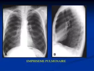
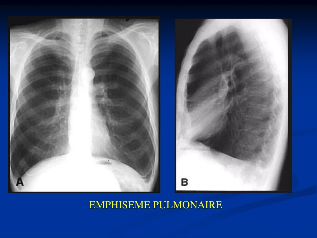
EMPHISEME PULMONAIRE. Radiologie– pneumonie au stafiloccoc. - distribution bilaterale - abces a paroi mince - pleuresie bilaterale - poche pleurale gauche. pneumatocel. Pneumonie avec bacililes enterics Gram negatives

E N D
Radiologie– pneumonie au stafiloccoc - distribution bilaterale- abces a paroi mince- pleuresie bilaterale- poche pleurale gauche pneumatocel
Pneumonie avec bacililesenterics Gram negatives Pseudomonas aeruginosa – piocyanic, Proteus mirabilis, Escherichia coli, Serratiamarcescens → imunodefficients Expectoration:verte (Pseudomonas), rouge (Serratia) Diagnostic:hemoculture Aspect en radiologie (Pseudomonas) ·aspect de bronhopneumonie - multiples abces· evolution recidivante· petites colections pleurales pneumonie (Pseudomonas) abcede lobe sup. gauche- image mixte hidro-aerienne au niveau orisontale
Aspecte radiologice - opacitati hiliobazale sub forma de cordoane liniare, intre care exista opacitati nodulare si reticulare - infiltrat neomogen (“barba pieptanata”) in triunghiul hiliobazal / perihilar / intercleidohilar - ex. dinamic: stergerea conturului si diminuarea contrastului in inspir profund TRIUNGHIUL DITTMAR RUPERT PNEUMONIE INTERSTITIALA: GRIPA
- acelaşi caz prezentat anterior; imagine hidro-aerică localizată în lobul superior drept- aspect dupa vomica (săgeată)
în interiorul ariei de condensare pulmonară se observă două imagini hidro-aerice suprapuse (aspect în gură de cuptor)
Trianghlebronchiectasique BRONCHIECTASIE BILATERALE: suprainfection
Patologie bronsica ULCERATII “NISE” PARIETALE BRONCHITE DEFORMANTE
ABCES PULMONAIR IN PHASE CRONIQUE “CHIRURGICALE”
Radiografii toracice - Cancerul masiv al hilului Massse hilaire droite Opacite hilaire gauche
Radiografii toracice - Pneumonia canceroasăopacitate de tip pneumonic lob inferior drept
- opacite homogene du hemitorace gauche - atelectasie poumon droit
1 2 pneumotorax totale1 – hipertransparence avec absence du dessin vasculaire2 - poumon colabe au hile pneumotorax totale gauche
Radioscopia: opacitate omogena, de intensitate ce variaza dupa cantitatea de lichid - image de pleuresie basale droite- opacite d’ intensite medium, a la limite superieure, flou, en menisc, concave au superieur
epanchement pleuraleopacite en menisc a la limite superieure concave en haut et dedans – dans les 2/3 inferieures du hemithorace gauche
opacite homogene du hemithorace droit; mediastinului pousse au contrelateral