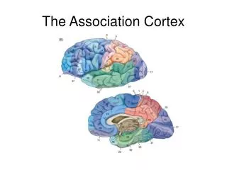
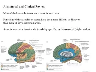
Anatomical and Clinical Review Most of the human brain cortex is association cortex. Functions of the association cortex
Anatomical and Clinical Review Most of the human brain cortex is association cortex. Functions of the association cortex have been more difficult to discover than those of any other brain areas. Association cortex is unimodal (modality specific) or heteromodal (higher order). Cerebral Cortex.

Anatomical and Clinical Review Most of the human brain cortex is association cortex. Functions of the association cortex
E N D
Presentation Transcript
Anatomical and Clinical Review Most of the human brain cortex is association cortex. Functions of the association cortex have been more difficult to discover than those of any other brain areas. Association cortex is unimodal (modality specific) or heteromodal (higher order).
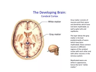
Cerebral Cortex • Brain’s most complex area with billions of neurons and trillions of synapses: the tissue responsible for mental activities • Consciousness • Perceives sensations • Commands skilled movements • Emotional awareness • Memory, thinking, language ability • Motivation • All “higher” mental functions
Types of Cerebral Cortex Neocortex • Newest in evolution • About 90% of total cortex in humans • 6 layers, most complex
Cerebral Cortex • Highly developed in humans, cetaceans, and primates • Makes up about 50% of total brain weight in humans • Surface area of 2.5 square feet in humans • 4.5 mm thick in precentral gyrus; 1.5 mm thick in visual cortex • Only 1/3 of surface area visible, 2/3 in banks of sulci • Surface of gyri and sulci • Estimated to contain about 15 billion neurons in humans
Histology of the Cerebral Cortex 1 • 2 main cell types are pyramidal and granule cells • Pyramidal cells have large apical dendrite and basal dendrites • Axon projects downward into subcortical white matter; may have collaterals • Pyramidal cell is the primary output neuron
Histology of Cerebral Cortex 2 • Pyramidal neurons are large and complex • Similar orientation • Process input from many sources
Histology of Cerebral Cortex 3:Dendritic Spines • Surfaces of dendrites covered with spines • Increase dendrite surface area for synapses • Faulty development of dendrites and spines seen in Down’s Syndrome • Dendrite complexity and spine numbers first increase postnatally and then decrease with old age
Dendritic Spines • Spines become more complex postnatally • Spine abnormalities occur in some conditions where mental performance is diminished Trisomy 21 Trisomy 13
Histology of Cerebral Cortex 3 • Granule (stellate) cells are interneurons • Short dendrites extending in all directions • Short axon projecting to adjacent pyramidal cells • Granule cells are expecially numerous in sensory and association cortex • Horizontal cells (layer I) and multiform cells (layer VI)
Functional Histology of the Cerebral Cortex • Neocortex has 6 layers designated I, II, III, IV, V, VI • Pyramidal cells predominate in layers III and V • Granule cells in layers II and IV
Types of Cortex • Cytoarchitecture varies in different areas • Number and size of cells • Thickness of layers
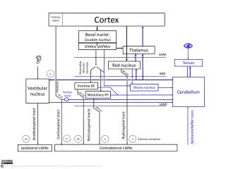
Cortical Columns • Functional units are cortical columns • Columns are vertically oriented groups of thousands of neurons in synaptic contact • Main input layer is layer IV which receives thalamic input • Thalamus is themain source of input to the cortex
Functional Histology • Layers V and VI = output • V to Basal ganglia, brainstem and spinal cord • VI to thalamus • Layers I, II, III = associative; projecting to other cortical areas • Layer IV = layer receiving inputs from thalamus and other cortical areas CC V VI
Cortical Connections 1 • Intracortical fibers • Association fibers • Commissural fibers • Subcortical fibers
Cortical Connections 2 • Intracortical fibers • short, project to nearby cortical areas • most from horizontal neurons in layer I • some from axon collaterals of pyramidal cells
Cortical Connections 3 • Association fibers • gyrus to gyrus and lobe to lobe in the same hemisphere • arcuate fibers connect adjacent gyri • long association fibers connect distant gyri • originate from pyramidal neurons in layers II and III
Cortical Connections 4 • Commissural fibers • connect homologous areas of the two hemispheres • Corpus callosum: rostrum, genu, trunk, splenium • rostrum & genu connect frontal lobes • trunk connects posterior frontal lobes, parietal lobes, and superior temporal lobe • splenium connects the occipital lobes • Originate with pyramidal neurons in layers II and III
Cortical Connections 5 • Anterior commissure connects the inferior and middle temporal gyri in opposite hemispheres; also olfactory connections • Posterior commissure carries fibers from the pretectal nuclei and other nearby neurons
Cortical Connections 6 • Projection fibers connect cortex with subcortical neurons • corticofugal/efferent, project from cortex • corticopetal/afferent, project to cortex • Corticofugal project to corpus striatum, brainstem, and spinal cord • Corticopetal projections arise mainly from the thalamus - the thalamic radiations • Internal capsule carries most of these connections
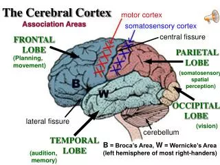
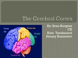
Functional Areas of Cerebral Cortex 1 • Anatomically the cortex is divided into 6 lobes: frontal, parietal, temporal, occipital, limbic and insular • Each lobe has several gyri • Functionally the cortex is divided into numbered areas first proposed by Brodmann in 1909 • Brodmann’s areas were described based on cytoarchitecture; later they were found to be functionally significant
Cortex cytoarchitecture Area 3 = postcentral gyrus Area 5 = superior parietal lobule Area 4 = frontal lobe primary motor cortex
Functional Areas of Cerebral Cortex 2 • Cytoarchitecture is based on the density and numbers of different cortical neurons and thickness of layers
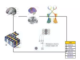
Functions include higher-order sensory processing, visual-spatial orientation, motor planning, language processing and production, determining socially appropriate behavior and “abstract thought.” Numerous subcortical structures are involved in these functions as well including thalamus, basal ganglia, and subcortical white matter. Complex functions of the brain are thought to involve both distributed networks of neurons and localized groups of neurons in specific cortical areas.
Unimodal sensory association cortex receives most input from primary sensory cortex of a specific modality and performs higher order sensory processing for that modality. Unimodal motor association cortex projects primarily to primary motor cortex and functions in formulating motor programs for complex actions involving multiple joints. Heteromodal association cortex has reciprocal connections with both motor and sensory association cortex of all modalities. Heteromodal assoc. cortex also has reciprocal connections with limbic cortex. This organization enables heteromodal assoc. cortex to perform the highest-order mental functions. This includes integration of abstract sensory and motor information from unimodal assoc. cortex, together with emotional and motivational influences provided by limbic cortex.
Principles of Cerebral Localization and Lateralization For over 100 years there have been 2 different theories about how the human brain is organized. One group supported the widespread network theory and the other group the localized functional regions theory. We now know that the brain has both types of functional organization. Localized regions carry out specific functions, but they do so through network interactions with many other regions of the brain. Focal brain lesions can cause specific deficits as will be seen in several examples such as aphasia and unilateral neglect, but most functions are also carried out by interconnected networks located in different brain regions, so false localization can occur in different injuries. Frontal lobe functions involve networks of neurons in different brain regions including frontal, parietal, and limbic cortices, thalamus, basal ganglia, cerebellum and brainstem.
Disconnection syndromes occur when fiber connections between different parts of a functional network are damaged. For example, if an injury damages white matter connections between visual cortex and the language processing areas in the adjacent association cortex, a patient may lose the ability to read. Hemispheric specialization is the tendency of some functions to be lateralized to the left or right hemisphere. One example is that the brain areas involved in language understanding and production are typically in the left hemisphere in humans. Handedness is another lateralized function. About 90% of humans are right- handed, so that performing tasks such as writing or closing buttons with the nondominant hand is very difficult. Evidence shows that skilled complex motor tasks for both right and left limbs are programmed mainly by the dominant hemisphere, so injury to the dominant hemisphere will lead to more severe motor deficits.
Language is lateralized in humans. The left hemisphere is dominant for language in over 95% of right-handers and 60-70% of left-handers. Many left-handers have significant bilateral language function. After a left hemisphere injury left handers tend to recover language more quickly than right-handers. Nondominant hemisphere is specialized for certain nonverbal functions. Complex visual-spatial skills, imparting emotional significant to events and language, and music perception are typically nondominant hemisphere functions.
The Dominant Hemisphere: Language Processing and Related Functions Wernicke’s area (22) functions to enable particular sequences of sounds to be identified and comprehended as meaningful words. Parts of adjacent areas 37, 39, and 40 are often included because lesions of these areas can produce Wernicke’s aphasia. Speech production function is carried out by Broca’s area (44, 45). Adjacent parts of cortical areas 9, 46, 47 or parts of even more distant areas 6, 8, 10 are sometimes also included. Ability to hear a word and then repeat it aloud requires the connecting path from Wernicke’s area to Broca’s area be intact. This path is called the arcuate fasciculus.
Sounds are converted into words by Wernicke’s area and then neural representations of words are converted back into sounds by Broca’s area. Broca’s and Wernicke’s areas do not carry out their functions alone. They have reciprocal connections with a large network of cortical areas that are also involved in language functions. Broca’s network connections: prefrontal cortex, premotor cortex, supplementary motor area. Function in speech formulation, planning and grammatical structure. Wernicke’s network connections: supramarginal gyrus, angular gyrus, and temporal lobe area 37. Function in language comprehension and lexicon for converting sounds to words and meanings.
The angular gyrus is especially important in reading. Visual inputs go to visual cortex and then are processed in visual association cortex, then to the angular gyrus and on to Wernicke’s area. Connections through the corpus callosum to the nondominant hemisphere function in language processing. Damage to nondominant hemisphere often produces difficulty judging the emotional tone of voice or may have problems producing speech with the correct emotional content. These connections can also be important if the dominant hemisphere is damaged and the nondominant hemisphere is needed to carry out language function (often requiring therapy/training). Lesions of thalamus, basal ganglia or adjacent white matter in the dominant hemisphere can produce aphasia that can be mistaken for a cortical lesion.
Aphasia A defect in language processing caused by dysfunction of the dominant cerebral hemisphere. Aphasia can be confused with other brain disorders.
The most common cause of acute onset aphasia is cerebral infarct.
Broca’s Aphasia Most common cause is stroke in the superior division of the middle cerebral artery. Most obvious symptom is decreased fluency in which phrase length is short (less than 5 words); more content words (nouns) than function words (prepositions and articles). Prosody, the normal melodious intonation of speech that conveys meaning, is lacking. Naming difficulties are common; comprehension is relatively intact.
Wernicke’s Aphasia Most commonly caused by stroke in the inferior division of the middle cerebral artery. Markedly impaired comprehension of language; may not be able to follow any instructions. Spontaneous speech has normal fluency, prosody and grammatical structure but is empty, meaningless, and full of nonsensical paraphasic errors. Patients are often unaware they have any language deficit. Anger or paranoid behavior is common.
Functional Areas of Cerebral Cortex Frontal Lobe Primary motor cortex, precentral gyrus, area 4 Premotor cortex, area 6, motor programs, apraxia (inability to perform movements in absence of paralysis) Frontal eye field, area 8 Supplementary motor cortex, parts of areas 6 & 8, programming for complex movements Prefrontal cortex, areas 9, 10, 11, 12, 32, 46, and 47, orbitofrontal area functions in visceral and emotional activities; dorsolateral area functions in intellectual activities such as planning, judgement, problem solving and conceptualizing 6. Broca’s area, 44 & 45, production of speech
Parietal Lobe Primary somatosensory cortex, postcentral gyrus, areas 1, 2, 3 Secondary somatosensory cortex, insula, posterior part of area 43 Primary gustatory cortex, anterior part of area 43 Parietal association cortex, areas 5, 7, 39, 40
Parietal Neglect SyndromeClinical Illustration • Failure to recognize side of body contralateral to injury • May not bathe contralateral side of body or shave contralateral side of face • Deny own limbs • Objects in contralateral visual field ignored
Temporal Lobe Primary auditory cortex, superior temporal gyrus, areas 41, 42 Auditory association cortex, Wernicke’s area 22, Wernicke’s aphasia Limbic cortex, areas 20,21, 27,28,29,30, 34,36,38; functions in emotions, complex behaviors and memory; amnesia, Alzheimer’s disease, prosopagnosia (20, 21)
Occipital Lobe Primary visual cortex, upper and lower banks of calcarine fissure, area 17 Visual association cortex, areas 18, 19; complex processing for color, movement, direction, visual interpretation; lesion can cause visual agnosia