
Digestive System
Digestive System. Digestive Tract. Also called alimentary canal Hollow tube roughly 8 meters in length. Structure of the Wall. Lumen - hollow center of tube Mucosa - epithelial layer with mucous-secreting cells

Digestive System
E N D
Presentation Transcript
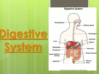
Digestive Tract • Also called alimentary canal • Hollow tube roughly 8 meters in length
Structure of the Wall • Lumen - hollow center of tube • Mucosa - epithelial layer with mucous-secreting cells • Submucosa - connective tissue layer rich with blood vessels, lymphatic vessels, and nerves • Muscular layer - smooth muscle layer • Circular- adjust lumen diameter • Longitudinal- adjust tract length • Serosa - outermost layer; secretes serous fluid
Types of Digestion • Mechanical • Physical breakdown of food into smaller pieces • Chemical • Breakdown of food molecules into more simple molecules by enzymes
Mouth • Receives food • Pushes into the remaining digestive tract • Includes: lips, teeth, cheeks, tongue, and palate • Two cavities: • 1) Oral cavity • Space between palate and tongue • 2) Vestibule • Cavity between teeth and lips and cheeks
1. 7. Exit Slip 2. 8. 9. 3. 10. 4. 11. 5. 6.
Cheek and Lips • Cheeks • Contain muscles used during chewing • Stratified squamous epithelial tissue inside • Lips • Highly mobile skeletal muscles rich in sensory receptors • Aid in sensing temperature and texture of food
Tongue • Functions: • Keeps food underneath teeth • Mixes food with saliva • Moves bolus to the back of the mouth during swallowing • Muscular structure covered with mucous membrane • Root is attached to hyoid bone • Attached to floor of mouth by frenulum • Papillae provide surface friction and contain taste buds
Palate • Forms roof of oral cavity • Hard palate • Anterior portion • Soft palate • Posterior portion; includes uvula • Soft palate raises during swallowing to close off nasal cavity
Tonsils • Masses of lymphatic tissue • Lingual • On the tongue • Pharyngeal • Posterior wall of pharynx; also called adenoids • Palatine • Back of mouth on either side of tongue; associated with palate
Teeth • Two sets: • 1) Primary - 20 teeth • Lost/shed, nonpermanent • 2) Secondary - 32 teeth • Permanent, come in after primary teeth are lost/shed • Function: • Begin mechanical digestion of food
Anatomy of Tooth • Crown - section above gingiva (gum) • Root -section below gingiva • Enamel - outer covering on crown • Dentin - bone-like substance that fills most of the tooth • Pulp cavity - connective tissue that contains blood vessels and nerves • Root canal - tubular extension that brings blood vessel and nerve to the pulp cavity • Cementum and periodontal ligament - hold tooth in alveolar process of jaw bone
Salivary Glands • Functions: • Moistens food • Binds food together • Dissolves food (so it can be tasted) • Cleanses mouth and teeth • Begins digestion of carbohydrates • Two types of cells • 1) Serous cells • Secretes serous fluid with enzyme amylase • 2) Mucous cells • Secretes mucous
Salivary Glands (cont) • 3 Types: • 1) Parotid glands: • Largest glands; anterior and inferior to ear; secrete watery saliva rich in amylase • 2) Submandibular: • Located in floor of mouth just inside lower jaw • 3) Sublingual: • Smallest glands; inferior to tongue; secrete saliva in mucous concentration
Squid Pharynx • Cavity located posterior to oral cavity • Provides connection to larynx and esophagus • Three parts: • 1) Nasopharynx- upper potion connecting to nasal cavity • 2) Oropharynx- middle section posterior to palate • 3) Laryngopharynx- lower portion posterior to larynx opening; leads to esophagus
Swallowing Swallowing Action • Bolus stimulates sensory receptors in pharyngeal opening • Soft palate raises- closes nasal cavity • Larynx elevates; epiglottis closes off larynx • Tongue presses against palate • Longitudinal muscle pull pharynx towards food • Muscles relax near esophagus to open the tube • Peristalsis moves food into esophagus
Esophagus • Hollow collapsible tube • Move food from pharynx to stomach • Passes through diaphragm in opening called esophageal hiatus • When food reaches opening of stomach, lower esophageal sphincter opens Neck
Stomach • J-shaped pouch in abdomen • Holds about 1 liter of food • Functions: • Mix food with gastric juices • Begin protein digestion • Responsible for limited absorption • Moves food into small intestine Squid Continues
Stomach (cont) • Rugae • Thick folds of mucosa and submucosa allow for expansion of stomach wall • Regions of the stomach • Cardiac - portion near esophagus • Fundic - portion lateral to cardiac where stomach ballons • Body - main portion of stomach between cardiac and pyloric • Pyloric - portion near opening to duodenum • Pyloric Sphincter - thick muscle band controlling entrance into duodenum
Gastric Secretions • Mucosa is studded with gastric pits • Gastric pits are the opening to gastric glands • Gastric glands have three types of secreting cells: • 1) Mucous cells - secrete mucous; helps prevent stomach from digesting itself • 2) Chief cells - secrete pepsinogen • 3) Parietal cells - secrete HCl and intrinsic factor
Gastric secretions (cont) • As food enters stomach, mixing actions occur to breakdown food into chyme • Gastric juices are added • HCl creates acidic environment • Shortens (activates) pepsinogen and makes it pepsin • Helps with vitamin B12 absorption
Gastric secretions (cont) • Limited absorption of the following occur: • Water • Salts • Alcohol • Lipid-soluble drugs • Chyme is moved to pyloric sphincter and pushed through
Control of Gastric Secretions • Digestion is controlled by medulla oblongata • Parasympathetic NS: • Increases gastric secretions • Sympathetic NS: • Decreases gastric secretions • Hormones: • Gastrin - stimulates production of gastric juices • Cholecystokinin - released when small intestine fills with food; decreases gastric motility
Review QuizMouth->Stomach • Contains rugae(folds)? 2. Contains lower sphincter and opens to stomach? 3. Mixes food with gastric juices? 4. Provides connection to larynx and esophagus? 5. Begins mechanical digestion of food? • Stomach • Esophagus • Stomach • Pharynx • Teeth
Pancreas • Has endocrine and exocrine function (Ch 11!) • Nestled in C-shaped curve of duodenum • Pancreatic acinar cells • Secrete pancreatic juices • Clustered around tubes that eventually empty into pancreatic duct • Pancreatic duct run the length of the pancreas • Empties the juice into the duodenum • Hepatopancreatic sphincter • Controls emptying
Pancreatic Enzymes • Carbohydrates: • Pancreatic amylase • Breaks polysaccharides into dissaccharides • Lipids: • Pancreatic lipase • Breaks fats into glycerol and fatty acids • Nucleic Acids: • Nucleases • Breaks nucleic acids into nucleotides
Pancreatic Enzymes (cont) • Protein: • 3 enzymes (break them down into amino acids) • Trypsin • Chymotrypsin • Carboxypeptidase • Stored in zymogen granules in inactive forms • Ex: Trypsin’s inactive form is trypsinogen and is activated by enterokinase which is secreted by mucosa of duodenum
Control of Pancreatic Juices and Enzymes • Parasympathetic NS control: • Stimulate release of pancreatic juices • Acidic chyme: • Stimulates release of secretin into the bloodstream • Stimulates release of pancreatic juice high in bicarbonate ions • Chyme high in protein and fat • Stimulates release of cholecystokinin into bloodstream • Stimulates release of pancreatic juice high in digestive enzymes
Liver • Functions: • Controlling carbohydrate metabolism • Lipid metabolism • Protein metabolism • Storage • Blood filtering • Detoxification • Secretion of bile
Liver Structure • Connective tissue divides liver intolarger right lobe and smaller left lobe • Liver is further divided into lobules • Hepatic cells radiate around a central vein • Spaces between the hepatic cells are called hepatic sinusoids
Liver Structure (cont) • Blood from digestive track enters sinusoids from hepatic portal vein • Kupffer cells • Large macrophages (filter out pathogens from sinusoids) • Hepatic cells • Take out excess nutrients • Blood enters central vein and continues on its path back to the heart
What is Bile? • Yellowish-green liquid secreted by liver cells • Includes: • Bile salts, bile pigments, cholesterol, and electrolytes
What does bile do? • Emulsification • Breaks fats globules into smaller droplets • Smaller droplets are easier for lipases to digest • Enhances absorption of fatty acids, cholesterol, and fat-soluble vitamins A, D, E, and K
Bile (cont) • Sequence of travel: • Hepatic cells → Bile canaliculi → Bile Ductules → Bile duct → Hepatic duct → Common hepatic duct → Hepatopancreatic sphincter → Duodenum
Gallbladder • Between meals, bile up in common hepatic duct and into the cystic duct that attaches to it • Bile is backed up into gallbladder that is attached to the cystic duct • Gallbladder also absorbs excess water in bile therefore concentrating it
Control of Bile Release • Chyme high in protein and fat • Stimulates release of cholecystokinin into bloodstream which stimulates release of bile • Where peristalsis reaches hepatopancreatic sphincter, it relaxes and bile squirts into duodenum
Small Intestine Structure • Three parts: • 1) Duodenum • First part after stomach; forms a C-shape • 2) Jejunum • More active than ileum • 3) Ileum • Leads to large intestine


