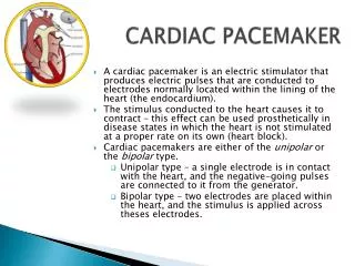
Artificial pacemaker and anesthesia
Artificial pacemaker and anesthesia. Presenter: Abraham sonny Bala chandran Moderator:Rashmi Ramachandran. www.anaesthesia.co.in anaesthesia.co.in@gmail.com. Preop assessment . Device assessment Patient evaluation and preparation. Preop assessment-Device.

Artificial pacemaker and anesthesia
E N D
Presentation Transcript
Artificial pacemaker and anesthesia Presenter: Abraham sonny Bala chandran Moderator:Rashmi Ramachandran www.anaesthesia.co.in anaesthesia.co.in@gmail.com
Preop assessment • Device assessment • Patient evaluation and preparation
Preop assessment-Device • Device assessment: • Type of device • Indication of device • Pacemaker mode with its special function • Device functioning status
Preop assessment-Device • Clinical history and physical examination • Manufacturers ID card / CXR to identify type of device • 12 lead ECG with and without magnet • Assessment of device functioning
Preop assessment-Patient • Patient level of dependency to device • Intraop factors interfering the device function • Consideration of reprogramming • Investigations
Preop assessment-Patient • History and evaluation of disease that mandated pacemaker • Continue antiarrythmic drug and other cardiac drugs as mandated • Consider Electromagnetic and Mechanical Interference (EMI)
Conditions warrant pacemaker reprogramming • Pacemaker dependent patients • Major procedure in abdomen and chest • Rate adaptive feature of pacemakers • Suspension of antitachycardia function • Surgical procedures with high chances of EMI
Electromagnetic and mechanical interference and pacemaker • Radio wave frequency between 0 -10 9 Hz and microwaves frequency 109 – 10 11 Hz cause EMI • Shielding of pulse generator, bipolar leads, and filtering of incoming signals offers some protection against EMI • Frequencies 5 -100 Hz overlap with intracardiac signals
Electromagnetic and mechanical interference and pacemaker • High probability of EMI • Electrocautery use • Lithotripsy • ECT • Radiation therapy • MRI • Orthopaedic procedures
Electromagnetic and mechanical interference and pacemaker • EMI effects on pacemaker: • Asynchronous pacing • Pacemaker inhibition or triggering • Resetting or reprogramming pacemaker • Damage to electronic circuits • Burn injury to myocardium • Inappropriate activation of rate adaptive feature and anti tachycardia therapy
Intraoperative management • Monitoring • Anesthetic technique • Measures to prevent device malfunctioning • Pacemaker failure trouble shooting • Preparation for temporary pacemaker and defibrillation
Monitoring • Continuous ECG • Signs of peripheral perfusion • Invasive monitoring Cardiac status Type of surgery Pacemaker failure
Anesthetic technique • Tailored to cardiovascular disease status • Anesthetics and pacing threshold • Succinylcholine and pacemaker
Electrocautery and pacemaker Uses radiofrequency of 300- 500 kHz • May affect pacemaker related to distance and orientation with the device
Electrocautery and pacemaker • Unipolar vs Bipolar vs Ultrasonic scalpel • Distance to pulse generator • Unipolar return pad • Lowest energy possible in short bursts
Magnet and pacemaker • Activates magnetic reed switch • Model dependent behaviour with magnet
Pacemaker magnet behaviour • Asynchronous pacing – most common • No apparent rate or rhythm change • Brief asynchronous pacing and then return to programmed value • Continuous or transient loss of pacing
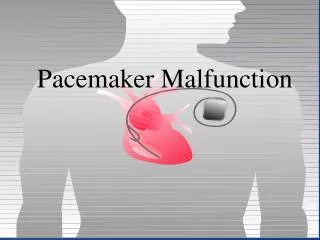
Pacemaker failure • Recognise • Assess haemodynamic status • Identify the cause • Manage
Pace maker failure • Failure to pace • Failure to capture • Undersensing / failure to sense • Oversensing
Failure to pace • Loss of pacing artifact in ECG in the absence of spontaneous QRS wave
Failure to capture • Absence of ‘P’or ‘QRS’ wave following atrial or ventricular artifact in ECG
Under sensing • Pacing artifact in the middle of spontaneous ‘P’ or ‘QRS’ wave -no change in automatic interval
Factors increasing pacemaker threshold • Myocardial ischaemia/infarct • Electrolyte disturbance • Acidosis or alkalosis • Hypoxia or hypercapnia • Abnormal antiarrythmic drug level especially Class I and beta blockers
Defibrillation • Defibrillation • Discontinue any obvious source of EMI • Restore antitachycardia function if available in device and BP stable • Pad or paddle position • Defibrillation and cardioversion dose • Post shock interrogation/ reprogramming
MRI and pacemaker • Torque effect on pulse generator • Magnetic field gradient closes magnetic reed switch • MRI produces radiofrequency waves of 30- 3000Hz
MRI and pacemaker • Latest models has less chance of torque effect • RF waves cause inappropriate pacing • Heat energy through leads- burn injury • MRI generally considered contraindicated in patients with pacemakers
MRI and pacemaker • Sommer et al in a prospective study assessed effects of MRI on pacemaker 82 patients undergoing extrathoracic site MRI.In this study they found no pacemaker inhibiition or arrythmia induction or any other serious injury during or after procedure. Hence they concluded MRI is safe and can be performed under due precautions - Circulation 2006; 114(2): 1285
MRI and pacemaker • Nazarian et al studied 55 patients (31 with pacemakers and 24 with AICD) undergoing cardiac and non cardiac MRI and found no significant adverse effects. They also concluded MRI can be safely performed in these patients considering the benefits in these patients - Circulation 2006; 114 (12):1277
MRI and pacemaker • Both these above studies were done under ECG and pulse oximetry monitoring and after appropriate reprogramming of the cardiac rhythm management device. Hence recommended these precautions to be followed during the procedure and MRI can be safely done in patients with CRMD.
Lithotripsy and pacemaker • Pacemaker circuitry damage • Focal point of beam directed away from generator • Atrial pacing and rate adaptive function need to be disabled • Contraindicated in patients with piezoelectric circuit implanted in abdominal wall
Lithotripsy and pacemaker • Albers DD et al found that lithotripsy can be performed safely done after appropriate reprogramming of the CRMD provided focus of beam is 5 cms away from the device. This study was done on 20 patients - J Endourol 1995; 9(4): 3011
Lithotripsy and pacemaker • Asroff et al described a case report of 75 yr male with pacemaker safely undergoing lithotripsy after inactivating rate adaptive function of device. They also suggested pacemaker generator not be located in the abdominal wall - J Endourol 1997; 157(3) : 189
Electroconvulsive therapy & pacemaker • Little current pass through heart • Seizures may cause inappropriate activation of AICD and rate adaptive feature of pacemaker • Other features may left intact • In patients with inactivated AICD be prepared for external defibrillation if VT/VF occurs
Automatic Implanted Cardioverter Defibrillator and anesthesia • Pacing and shocking function • Precautions similar to pacemaker • Deactivate cardioversion and defibrillation function of AICD
Automatic Implanted Cardioverter Defibrillator and anesthesia • Magnet and ICD: • Pacemaker function is rarely altered • Suspends anti tachyarrythmia therapy • Some ICD have no magnet response • Most are permanently disabled • Hence recommended not to be used in patients with ICD
Automatic Implanted Cardioverter Defibrillator and anesthesia • External defibrillation in patients with AICD • Apply external defibrillator pad before surgery • Remove EMI source • Reenable antitachycardia function • If above measures fails provide external defibrillation with precaution as for pacemakers
Postoperative care • Monitoring • Device interrogation • Reprogramming
Summary • Pacemaker and AICD implanted patients undergoing elective surgery: • Contact pacemaker or AICD clinic or manufacturer • Reprogram the device function in selective group of patients
Summary • Pacemaker and AICD implanted patients undergoing elective surgery: • Monitoring and anesthesia technique with due considerations to patients CVS status • Avoid electrocautery use. If necessary consider use of bipolar or harmonic scalpel • Be ready for alternate mode of pacemaker and defibrillator if necessity arises
Summary • Pacemaker and AICD implanted patients undergoing emergency surgery: • Try get information regarding pacemaker or AICD at earliest possible or if time permits • Initiate invasive arterial pressure monitoring in addition to standard monitoring
Summary • Pacemaker and AICD implanted patients undergoing emergency surgery: • If pacemaker failure is associated with haemodynamic instability consider magnet application • Consider magnet application with AICD only when EMI source cannot stopped and it is associated with haemodyanamically destabilising inappropriate shocks • Interrogate device following either elective or emergency surgery at the earliest possible and consider appropriate resetting if required
Thank you www.anaesthesia.co.in anaesthesia.co.in@gmail.com