Chapter 6 The Skeletal System: The Framework
Chapter 6 The Skeletal System: The Framework. Multimedia Directory. Slide 40 Joint Classification Animation Slide 41 Joint Movement Animation Slide 45 Ankle Dorsiflexion/Extension Animation Slide 46 Ankle Inversion/Eversion Animation Slide 47 Elbow Pronation/Supination Animation

Chapter 6 The Skeletal System: The Framework
E N D
Presentation Transcript
Chapter 6 The Skeletal System: The Framework
Multimedia Directory Slide 40 Joint Classification Animation Slide 41 Joint Movement Animation Slide 45 Ankle Dorsiflexion/Extension Animation Slide 46 Ankle Inversion/Eversion Animation Slide 47 Elbow Pronation/Supination Animation Slide 48 Elbow Flexion/Extension Animation Slide 49 Hand Opposition Animation Slide 50 Humerus Adduction/Abduction Animation Slide 51 Humerus Circumduction Animation Slide 52 Humerus Rotation Animation Slide 53 Wrist Circumduction Animation Slide 54 Wrist Flexion/Extension Animation
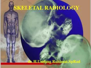
Multimedia Directory (cont’d) Slide 60 Skeletal System Exercise Slide 61 Skull Bones Exercise Slide 92 Bone Healing Animation Slide 97 Osteoporosis Video Slide 98 Arthritis Video Slide 123 Radiologic Technology Video
Introduction Skeletal system provides support and allows us to move Bones (also called osseous tissue) that make up skeleton protects soft body parts, produces blood cells, and acts as storage unit for minerals and fat There are 206 bones in adult skeleton, along with cartilage, ligaments, and joints
Learning Objectives Describe the functions of the skeletal system Identify and describe the anatomy and physiology of bone Locate and describe the various bones within the body
Learning Objectives (cont’d) Differentiate between bone, cartilage, ligaments, and tendons Locate and describe the various joints and types of movement of the body Explain common diseases and disorders of the skeletal system
Pronunciation Guide Appendicular skeleton Arthritis Articulation Axial skeleton Cancellous bone Diaphysis Epiphyseal plate Epiphysis Hemopoiesis Medullary cavity (app en DIK yoo lahr SKELL eh ton) (ahr THRYE tiss) (AHR tick you lay shun) (AK see al SKELL eh ton) (CAN cell us) (dye AFF ih siss) (eh piff ih SEE al) (eh PIFF ih siss) (HEME ah poy ee sus) (MED uh lair ee) Click on the megaphone icon before each item to hear the pronunciation.
Pronunciation Guide (cont’d) Osseous tissue Ossification Osteoarthritis Osteocytes Osteons Periosteum Synovial fluid Trabeculae Vertebrae (OSS see us) (OSS siff ih cay shun) (OSS tee oh ahr THRYE tiss) (OSS tee oh site) (OSS tee ons) (pair ee OSS tee um) (sin OH vee al) (tra BECK you la) (VER the bray) Click on the megaphone icon before each item to hear the pronunciation.
Bones Are primary components of skeleton Although composed of non-living minerals such as calcium and phosphorous, bones are very much alive, constantly building and repairing themselves Word ‘bone’ comes from Greek meaning “dried up body”
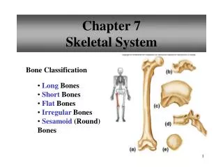
General Bone Classifications Classified according to their shape Long bones: longer than they are wide and can be found in arms and legs Short bones: fairly equal in width and length and found mostly in wrists and ankles Flat bones: thinner and can be either flat or curved; can be plate-like in nature and would include skull, ribs, and sternum (breast bone) Irregular bones: like parts of jigsaw puzzle, odd in shape, and include hip bone and vertebrae
Bone Anatomy Periosteum Tough and fibrous connective tissue covering bone Contains blood vessels which transport blood and nutrients to nurture bone cells Also contains lymph vessels and nerves Acts as anchor point for ligaments and tendons
Bone Anatomy (cont’d) Epiphysis and diaphysis Epiphysis: formed by increase in size of both ends of long bone Diaphysis: region running between two epiphyses; hollow area called medullary cavity acts as storage area for bone marrow Bone marrow Yellow marrow: has high fat content; can convert to red marrow in an emergency Red marrow: produces red blood cells
Bone Tissue Compact bone Dense, hard tissue that composes shafts of long bones and forms outer layer of other bone types Tightly packed material within tissue makes for dense and strong structure Material forms microscopic, cylindrical shaped units called osteons, or Haversian systems
Bone Tissue (cont’d) Each units has mature bone cells (osteocytes) forming concentric circles around blood vessels Area around osteocyte is filled with protein fibers, calcium, and other minerals Osteons run parallel to each other with blood vessels literally connecting with them to ensure sufficient oxygen and nutrients for bone cell
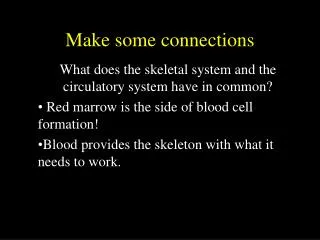
Bone Tissue (cont’d) Spongy (cancellous) bone Arranged in bars and plates called trabeculae Irregular holes between trabeculae make bone lighter in weight and provide space for red bone marrow, which produces red blood cells Holes give bone spongy appearance
Surface Structure of Bones Bone is not perfectly smooth; has variety of projections, bumps, and depressions Projections act as points of attachment for muscles, ligaments, or tendons Grooves and depressions act as pathways for nerves and blood vessels Projecting structures and depressions can work together as joining or articulation points to form joints such as ball and socket joint in hip
Bone Growth and Repair Ossification: describes formation of bone in body Bones grow longitudinally to develop height and horizontally (wider and thicker) so they can more efficiently support body weight and any other weight we support
Bone Growth and Repair (cont’d) Cell types involved in bone formation and growth Osteoprogenitor cells Osteoblasts Osteocytes Osteoclasts
Bone Growth and Repair (cont’d) Osteoprogenitor cells: non-specialized cells found in periosteum, endosteum, and central canal of compact bones; can turn into other types of cells as needed Osteoblasts: cells that actually form bones; arise from non-specialized osteoprogenitor cells and are cells that secrete a matrix of calcium with other minerals that give bone its typical characteristics
Bone Growth and Repair (cont’d) Osteocytes: considered mature bone cells that started as osteoblasts; osteoblasts surround themselves with matrix to then become mature osteocytes Osteoclasts: believed to originate from type of white blood cell called monocyte found in red bone marrow; job is to tear down bone material and help move calcium and phosphate into blood
Bone Growth and Repair (cont’d) Bone development begins when we are embryos through intramembranous and endochondral ossification Intramembranous ossification occurs when bone develops between two sheets composed of fibrous connective tissue
Bone Growth and Repair (cont’d) Cells from connective tissue turn into osteoblasts and form matrix while other osteoblasts create compact bone over surface of spongy bone; once matrix surrounds osteoblasts they become osteocytes, which is how bones of skull develop
Bone Growth and Repair (cont’d) Majority of bones form through endochondral ossification Shaped cartilage replaced by bone Periosteum surrounds diaphysis of cartilage bone as cartilage itself begins to break down Osteoblasts come into region and create spongy bone in area that is then referred to as primary ossification center
Bone Growth and Repair (cont’d) Other osteoblasts begin to form compact bone under periosteum; osteoclasts break down spongy bone of diaphysis to create medullary cavity
Bone Growth and Repair (cont’d) Epiphyseal plate (growth plate) After we are born, epiphysis on long bones continues to grow Plate is thin band of cartilage formed between primary and secondary ossification centers Plate exists as long as bones need to lengthen and widen; controlled by hormones, plate will eventually ossify and stop growth process
Pathology Connection: Osteoporosis As we age breakdown of bone becomes greater than formation of new bone (causing bone mass to gradually decrease) Bones become lighter and weaker, with holes in spongy bone becoming more prominent; weakened bones more prone to breakage; condition of decreasing bone density called osteoporosis
Pathology Connection: Osteoporosis (cont’d) Loss of bone mass can be slowed down by Increasing calcium (forms matrix of bone), fluoride, and vitamin D (helps body absorb ingested calcium from digestive tract) in diet, particularly in formative years Eliminating smoking and decreasing caffeine consumption (both aid in calcium depletion) Engaging in weight-bearing exercise Taking medications to increase bone mass (such as alendronate)
Cartilage Special form of dense connective tissue that can withstand fair amount of flexing, tension, and pressure Flexible part of nose and ears are cartilage Also makes flexible connection between bones, as between ribs and sternum, allowing chest flexion during deep breathing
Cartilage (cont’d) Acts as cushion between bones; articular cartilage located on ends of bones and acts as shock absorber, preventing ends from grinding together when you move In this location, small sacs, called bursa, secrete lubricant called synovial fluid Joints can still wear out and become inflamed despite all this protection, resulting in arthritis or osteoarthritis
Joints and Ligaments When two or more bones join together they form a joint or articulation Articulating joints held together, yet still movable; accomplished by special connective tissue called ligaments Ligaments: tough, whitish bands that connect from bone to bone and can withstand heavy stress Tendons: cord-like structures that attach muscle to bone
Joints and Ligaments (cont’d) Types of synovial joints Pivot joint: turnstile movement in neck and forearm Ball and socket joint: hip and shoulder; all forms of movement, including rotation Hinge joint: allow opening and closing movement in knees and elbows Gliding joint: wrists and ankles; provides sliding back and forth movement
Joints and Ligaments (cont’d) Types of synovial joints Saddle joint: shaped like saddle, found in thumb; can rock up and down or side to side Condyloid joint: oval shaped bone end fitting into elliptical cavity in other bone so there is movement from one plane to another but no rotation as found in fingers and toes Ellipsoidal joint: provide two axes of movement through same bone like joint formed at wrist with radius and ulna
Joint Classification Animation Click here to view an animation showing classifications of the joints. Back to Directory
Joint Movement Animation Click here to view an animation showing movement of the joints. Back to Directory
Movement Classifications Flexion: bending a joint and decreasing angle between involved bones Extension: straightening a joint Plantar flexion: pointing toes down Dorsiflexion: bending foot up toward body Abduction: moving away from body’s midline Adduction: moving toward midline of body
Movement Classifications (cont’d) Inversion: turning foot inward toward other foot Eversion: turning foot outward away from opposing foot Supination: turning hand palm up Pronation: turning hand palm down Circumduction: circular arm movement of a pitcher
Ankle Dorsiflexion/Extension Animation Click here to view an animation showing ankle dorsiflexion/extension. Back to Directory
Ankle Inversion/Eversion Animation Click here to view an animation showing ankle inversion/eversion. Back to Directory
Elbow Pronation/Supination Animation Click here to view an animation showing elbow pronation/supination. Back to Directory
Elbow Flexion/Extension Animation Click here to view an animation showing elbow flexion/extension. Back to Directory
Hand Opposition Animation Click here to view an animation showing hand opposition. Back to Directory
Humerus Adduction/Abduction Animation Click here to view an animation showing humerus adduction/abduction. Back to Directory