
Pair Distribution Function-Computed Tomography Scans
Pair Distribution Function-Computed Tomography Scans. “One of the most significant developments in X-ray micro tomography for almost 30 years” Prof Robert Cernik , Manchester’s School of Materials. What do we have now?. X-ray tomography

Pair Distribution Function-Computed Tomography Scans
E N D
Presentation Transcript
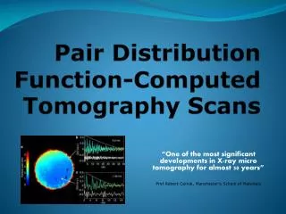
Pair Distribution Function-Computed Tomography Scans “One of the most significant developments in X-ray micro tomography for almost 30 years” Prof Robert Cernik, Manchester’s School of Materials
What do we have now? X-ray tomography This collects the X-rays transmitted by an object whilst it is rotated so that it an image of its 3D shape can be constructed. By comparing how the X-rays approach the object and how much they are blocked/transmitted we can figure out the shape of that face of the object. By rotating the sample we can construct similar images from every angle to create a 3D model of it. We can also find out the density of different parts of the object to create a density contrast image. This sometimes helps identify obvious abnormalities (e.g. an advanced tumour)
MRI Scans for MS Magnetic Resonance Imaging (MRI) Scans broadly use the tomography described above. If MS is suspected, a special contrast material (usually gadolinium) is injected just before the scan. Since it reacts to inflammation the reaction locations/areas of inflammationwill appear as light patches (a “contrast” to the mostly dark background). This shows where demyelination is occurring. Problems with MRIs: • 5% of people with MS do not have abnormalities that can be detected on an MRI (resulting in a false negative) • Some age-related damage looks like MS lesions (resulting in a false positive) • The test results take a few weeks, which has a worse impact the more severely you have the disease as you want treatment to begin as soon as possible to reverse and prevent damage.
What does the new scan do? It separates nanostructure signals from the object to see what the atoms are doing in each location. The new method allows us to more accurately describe the structure and chemistry of different parts of the object. This makes diagnosis much easier and more effective because doctors can be more certain of the nature and potency of what they are dealing with. For example, cancer or tumours can be identified when they are in their very early stages. It also allows us to image at the nanoscale, so we gain more detailed images and can model individual molecules we could not before.


