Download
1 / 16
190 likes | 461 Views
PLLA-PEG-TCH-labeled bioactive molecule nanofibers for tissue engineering. Haiyun Gao 1,2,3 , Jun Chen 1,3 , Beth Zhou 1,2,3 , Wen Zhong 3 and Malcolm Xing 1,2,4 1.Mechanical Engineering, Faculty of Engineering, 2.Manitoba Institute of Child Health,

E N D
PLLA-PEG-TCH-labeled bioactive molecule nanofibersfor tissue engineering Haiyun Gao1,2,3, Jun Chen1,3, Beth Zhou1,2,3, Wen Zhong3 and Malcolm Xing1,2,4 1.Mechanical Engineering, Faculty of Engineering, 2.Manitoba Institute of Child Health, 3.Textile Science, Faculty of Human Ecology and 4. Biochemistry and Medical Genetics, Faculty of Medicine, University of Manitoba.
Nanofibers S G Kumbar et al. Biomed. Mater. 3 (2008) • Fiber with nanodimensions • an extraordinarily high surface area to volume ratio • tunable optical emission • super paramagnetic behavior • Methods to fabricate nanofibers • Drawing • Template synthesis • Temperature-induced phase separation • Molecular self-assembly • Eletrospinning
Electrospinning strategy S G Kumbar et al. Biomed. Mater. 3 (2008) Advantages: • Simple instrument • Continuous process • Cost effective compared to other existing methods • Scalable • Ability to fabricate fiberdiameters from few nm to several microns • High efficiency for biomedicine application
Drug Loading in ElectrospunNanofibers (ENs) For my study: Drug loaded in ENs: tetracycline hydrochloride [TCH] a model antibiotic TCH • Wide applications for ENs: • Fltration, sensors, military protective clothing, photovoltaic devices, liquid-crystal display (LCD), ultra-light weight space craft materials, super-efficient and functional catalysts • Variety of biomedical applications: carriers for drug/therapeutic agent delivery, wound dressing materials and as porous three dimensional scaffolds for engineering various tissues such as skin, blood vessels, nerve, tendon, bone and cartilage
PLLA/PLLA-PEG-NH2ElectrospinningNanofibers PLLA PEG NH2 Preparation of solution: NaOH + tetrahydrofuran CH2Cl2 + trifluoroacetic acid (TFA) BTAC (surfactant) Blending electrospinning conditions: room temperature voltage of 22 kV flow rate of 7 mL/h distance of 12 cm between the needle tip and the collector PLLA/PLLA-PEG-NH2electrospunnanofiabers • Materials: • Poly(L-lactide) (PLLA) • Poly(ethylene glycol) (PEG) with functional group
TCH-loaded Nanofibers 3% w/w of TCH + 7.5% w/w PLLA/PLLA-PEG-NH2 + 5% w/w BTAC Emulsification and electrospinning Surface functionalization of electrospunnanofibers for the immobilization of proteins Model proteins: Two fluorescently tagged bovine serum albumins (BSAs) Red: Rhodamin-BSA Green: FITC-BSA HOW: emulsion electrospinning
Validity of the Immobilization Method NHS was coupled to the carboxyl groups of surface hydrolyzed ENS, resulting in the formation of an NHS ester, with the amide I band at 1646 cm−1 EGS was conjugated to the amino groups of water vapor-treated ENS to form an amide (1646 cm−1)
TCH-loaded Nanofibers Table 1 Sample names and specifications Morphology of the electrospunnanofibers (Scanning electron microscopy micrograph) H0-1 (641nm) H0-3 (608nm) H3-1 (740nm) H3-3 (780nm) Bar: 2 µm.
PLLA/PLLA-PEG-NH2nanofibersfunctionalized with both FITC-BSA and rhodamine-BSA Confocal images of PLLA/PLLA-PEG-NH2 nanofibers functionalized with both FITC-BSA and rhodamine-BSA. (A) Image showing FITC-BSA, (B) image showing rhodamine-BSA, and (C) merged A and B. Bar: 20 µm.
In Vitro Release of TCH and Antibiotic Susceptibility Test The encapsulation rate of TCH was 70% for ENSs without BSA conjugation (H3-1) and 30% for ENSs with two conjugated BSAs (H3-3).
Antibiotic Susceptibility Test Antibacterial tests of H3-1 and H3-3. (A) Day 1 H3-3 (left) and H3-1 (right); (B) day 2 H3-3 (left) and H3-1 (right); (C) day 3 H3-3 (left) and H3-1 (right); and (D) day 4 H3-1 (H3-3 was discarded). Bar: 5 mm.
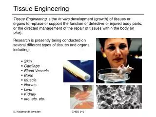
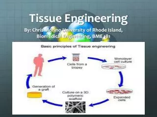
ENs in Tissue Engineering Tissue engineering? Bioresorbable and biocompatible materials native tissues Mimic /Replicate In one approach to open system implants, three-dimensional highly porous scaffolds composed of synthetic polymers serve as cell transplant devices. Langer, R. and J. P. Vacanti (1993). "Tissue Engineering." Science 260(5110): 920-926.
ENs in Tissue Engineering ENs Tunable porosity maintain cellular functionality cells exchange metabolites and nutrients with environment aid in the reconstruction of tissues maintaining tailored mechanical properties to protect the wound bed from collapse avoid mechanical mismatch between scaffolds and host tissues
Cell adhesion and proliferation on nanofibrous scaffold conjugate PDGF-BB/RGDS(promote cell attachment) ENs Immunofluorescence staining of human dermal fibroblasts on ENs of (A) PDGF and RGDS conjugated and (B) blank. Images were recorded by a confocal microscope. The same magnification was used for both pictures. Bar: 100 µm.
Conclusion Multifunctional ENSs were developed that incorporated the antibacterial agent TCH and were successfully surface functionalized with two different bioactive molecules. This novel material may have potential applications in wound care and tissue engineering.
ACKNOWLEDGEMENT Acknowledgement comes to the Institute of Textile Science, University of Manitoba, Manitoba Institute of Child Health and all my colleagues.