Tissue Engineering
Tissue Engineering. Tissue Engineering is the in vitro development (growth) of tissues or organs to replace or support the function of defective or injured body parts, or the directed management of the repair of tissues within the body ( in vivo ).

Tissue Engineering
E N D
Presentation Transcript
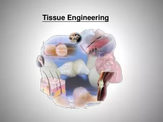
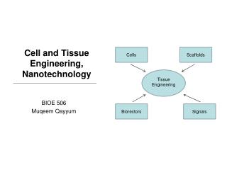
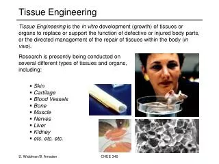
Tissue Engineering • Tissue Engineering is the in vitro development (growth) of tissues or organs to replace or support the function of defective or injured body parts, or the directed management of the repair of tissues within the body (in vivo). • Research is presently being conducted on several different types of tissues and organs, including: • Skin • Cartilage • Blood Vessels • Bone • Muscle • Nerves • Liver • Kidney • etc. etc. etc. CHEE 340
Tissue Organization • Before a tissue can be developed in vitro, first we must understand how tissues are organized. The basic tenet here is that: • “all tissues are comprised of • several levels of structural hierarchy” • These structural levels exist from the macroscopic level (centimeter range) all the way down the molecular level (nanometer range) • there can be as many as 7-10 distinct levels of structural organization in some tissues or organs CHEE 340
Organization of the Tendon CHEE 340
Organization of the Kidney CHEE 340
Functional Subunits • The smallest level at which the basic function of the tissue/organ is provided is called a “functional subunit”: • functional subunits are in the order of ~100 mm (whereas cells are of the order of ~10 mm) • each organ is comprised between 10-100 x 106 functional subunits • each functional subunit is comprised of a mixture of different cell types and extracellular matrix (ECM) molecules • Separation of the functional subunit into individual cohorts (i.e. cells and ECM) leads to a loss of tissue function. For this reason, this is the scale that tissue-engineering tries to reconstruct. • So, how can the functional subunit be built in vitro? CHEE 340
Microenvironment • Since cells are entirely responsible for synthesizing tissue constituents and assembly of the functional subunit, much attention is paid to the microenvironment surrounding the cell(s) of interest. • The microenvironment, which can be very different depending on the type of cell, is typically characterized by the following: • Cellularity • Cellular Communications • Local Chemical Environment • Local Geometry CHEE 340
Cellularity • Packing Density: • maximum theoretical packing density is about 1 x 10⁹ cells/cm3 • cell densities in tissues typically vary between 10 – 500 x 106 cells/cm3 • relates to about 100 - 500 cells per microenvironment (100 mm)3 • extreme cases, such as cartilage which has ~ 1 cell per (100 mm)3 • thus its microenvironment is essentially 1 cell plus associated ECM • Cellular Communication: • Cells communicate in three principal ways: • secretion of soluble signals • cell-to-cell contact • cell-ECM interactions • Cellular communication can affect all “cellular fate” processes (migration, replication, differentiation, apoptosis) and the method(s) of communication used depends, in part, on how the cells are packed within the tissue. CHEE 340
Cellular Communications • Soluble Signals: • includes small proteins such as growth factors and cytokines (15-20 kDa), steroids, hormones • bind to membrane receptors usually with high affinity (low binding constants: 10-100 pM) CHEE 340
Cellular Communications • Cell-to-Cell Contact: • some membrane receptors are adhesive molecules • adherent junctions and desmosomes • other serve to create junctions between adjacent cells allowing for direct cytoplasmic communication • gap junctions • 1.5-2 nm diameter and only allow transport of small molecules ~1 kDa CHEE 340
Cellular Communications • Cell-ECM Interactions: • ECM is multifunctional and also provides a substrate that cells can communicate through • since cells synthesize the ECM, they can modify the ECM to elicit specific cellular responses • cells possess several specialized receptors that allow for cell-ECM interactions • integrins, CD44, etc. • also a mechanism by with cells respond to external stimuli (“mechanical transducers”) CHEE 340
Chemical Environment • Oxygenation: • mammalian cells do not consume oxygen rapidly but uptake is large in comparison to the amount in blood or culture media • air-saturated aqueous media (37°C) contains only 21 mM O2 • mammalian cells consume O2 at rate of 0.05-0.5 mmol/106 cells/hour • cell cultures for tissue engineering have relatively large cell densities (106 cells/mL) which results in total O2 depletion in 0.4-4 hours! • concentration must be within a specific range since oxygenation affects a variety of physiological functions • low O2 concentration can retard growth • high O2 concentration can be inhibitory or toxic (oxidative stress) • Metabolism: • typically, there are no transport limitations for major nutrients although uptake rate depends on their local concentrations • glucose uptake rate: 0.1-0.5 mmol/106 cells/hour • amino acid uptake rate: 1.0-5.0 nmol/106 cells/hour CHEE 340
Local Geometry • Geometry of the microenvironment depends on the individual tissue: • needs to be re-created for proper tissue growth • two-dimensional layers or sheets • three-dimensional arrangements • transport issues • local geometry also affects how cells interact with the ECM • remember, the ECM serves as a substrate for cellular communications • For these reasons, considerable effort has been geared at creating artificial ECM’s (aka scaffolds) to provide the appropriate substrate to guide in vitro tissue growth and development. CHEE 340
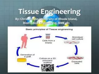
Tissue Engineering • General Paradigm SJ Shieh and JP Vacanti Surgery 137 (2005) 1-7
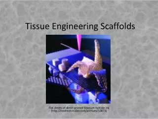
Tissue Engineering Scaffolds • Scaffold Materials: • synthetic polymers • poly(lactide) ,poly(lactide-co-glycolide), poly(caprolactone)…. • foams, hydrogels, fibres, thin films • natural polymers • collagen, elastin, fibrin, chitosan, alginate…. • fibres, hydrogels • ceramic • calcium phosphate based for bone tissue engineering • porous structures • permanent versus resorbable • degradation typically by hydrolysis (except for natural materials) • must match degradation rate with tissue growth • Chemical and Physical Modifications (synthetic materials): • attachment of growth factors, binding sites for integrins, etc. • nanoscale physical features CHEE 340
Tissue Engineering Scaffolds smooth muscle cells on unmodified poly(CL-LA) elastomer (L) and modified surface having bound peptide sequence(R) CHEE 340
Culturing of Cells • Types of Cell Culture • monolayer (adherent cells) • suspension (non-adherent cells) • three-dimensional (scaffolds or templates) CHEE 340
Culturing of Cells • Sterilization Methods • ultra-violet light, 70% ethanol, steam autoclave, gamma irradiation, ethylene oxide gas • Growth Conditions • simulate physiological environment • pH 7.4, 37°C, 5% CO2, 95% relative humidity • culture (growth) media replenished periodically • Culture (Growth) Media • appropriate chemical environment • pH, osmolality, ionic strength, buffering agents • appropriate nutritional environment • nutrients, amino acids, vitamins, minerals, growth factors, etc. CHEE 340
Cell Sources • Since the ultimate goal of tissue engineering is to develop replacement tissue (or organs) for individuals, the use of autologous cells would avoid any potential immunological complications. • Various classifications of cells used in tissue engineering applications: • primary cells • differentiated cells harvested from the patient (tissue biopsy) • low cellular yield (can only harvest so much) • potential age-related problems • passaged cells • serial expansion of primary cells (can increase population by 100-1000X) • tendency to either lose potency or de-differentiate with too many passages • stem cells • undifferentiated cells • self-renewal capability (unlimited?) • can differentiate into functional cell types • very rare CHEE 340
Stem Cells • Stem cells naturally exist in essentially all tissues (especially those that rapidly proliferate or remodel) and are present in the circulation. • There are two predominant lineages of stem cells: • mesenchymal • give rise to connective tissues (bone, cartilage, etc.) • although found in some tissues, typically isolated from bone marrow • hematopoietic • give rise to blood cells and lymphocytes • isolated from bone marrow, blood (umbilical cord) • Stem cells are rare; bone marrow typically has: • a single mesenchymal stem cell for every 1,000,000 myeloid cells • a single hematopoietic stem cell for every 100,000 myeloid cells CHEE 340
Stem Cells (Mesenchymal) CHEE 340
Stem Cells (Hematopoietic) CHEE 340
Proliferation versus Commitment Proliferation Commitment or Differentiation Clonal Succession Stem Cell Deterministic or Stochastic Succession CHEE 340
Stem Cells • Identification • Stem cells are identified by the expression of specific antigens on their surface, for example: • hematopoietic stem cells express CD45, CD34 and CD14 • mesenchymal stem cells do not express these markers (i.e. CD34-, CD45-, CD14-) • Selective separation of positive marker cells (in a mixed cell population) can be done by several techniques (e.g. immunomagnetic methods). • Characterization and Commitment • The most common approach to characterize multi-lineage- or single lineage-committed stem cells is through colony-forming assays: • cells grown under culture conditions that promote their proliferation and differentiation • the clonal progeny of a single progenitor cell stay together to form a new colony of mature cells • colony-forming assays are used to: • characterize stem cells from different sources (e.g. BM, umbilical cord blood) • investigate responses to growth factors, cytokines and other drugs • expansion, commitment, etc. • quality control for collection, processing and cryopreservation CHEE 340
Colony-Forming Units (CFUs) CHEE 340
Scale Up • The conditions of the in vivo microenvironment are a fine balance between biological dynamics and the physiochemical processes that constrain them. Thus, the design of cell and tissue culture devices must be such that this balance is maintained down to about 100 mm – the size of the tissue microenvironment. • Several important design challenges: • mass transfer (delivery and removal) • fluid flow CHEE 340
Mass Transfer • The importance of mass transfer in tissue and cellular function is often overlooked. The diffusional penetration lengths over physiological time scales are surprisingly short and constrain the in vivo architecture of tissues and organs. • Similar constraints are faces with the construction of cell culture devices and it may be difficult to provide the appropriate mass-transfer rate into a cell bed of physiological cell density. • For any nutrient (O2, glucose, growth factor, etc.), there are two primary concerns for appropriate delivery: • provided at physiological concentrations • provided at the same rate it is consumed CHEE 340
C t= q X Mass Transfer Can estimate the time it takes to deplete a nutrient from the media using the following relation: t – time until total depletion [hours] C – concentration of nutrient [mM] q – specific nutrient consumption rate [mmol/cell/hour] X – number of viable cells per unit volume [cells/mL] The product (q X) is the total nutrient consumption rate for the particular system and this rate must be balanced with the total delivery rate to ensure proper cellular function. An imbalance between delivery and consumption will alter the local nutrient concentration which can have adverse affects on cellular function. • too high or too low can be inhibitory or even toxic CHEE 340
Mass Transfer Oxygen • physiological concentration: 5-30% of saturation in air, which is ~ 0.2 mM • specific uptake rate: 0.05 – 1 mmol/106 cells/hour Primary Nutrients (glucose) • physiological concentration: mM range • specific uptake rate: 0.05 – 0.1 mmol/106 cells/hour Secondary Nutrients (amino acids, growth factors) • physiological concentration: nM – mM range • specific uptake rate: 0.01 – 1.0 nmol/106 cells/hour Waste Products (lactic acid, ammonia) • physiological concentration: negligible • specific production rate: 0.01 – 0.2 mmol/106 cells/hour CHEE 340
Fluid Flow • The circulatory system provides blood flow to all of the microenvironments of the body. Overall, the perfusion rate in humans is about 5 L/min/person. This is roughly equivalent to 50 – 400 mL/min/106 cells: • different depending on the metabolic activity of the specific cell type • very low in magnitude compared to fermenters • such low flow can lead to additional problems such as surface-tension effects and capillary action • Fluid flow not only provides the delivery of nutrients (dissolved gases, glucose, growth factors, etc.) but also serves to remove inhibitory waste products, cytokines and degenerative enzymes: • waste products of metabolism: • carbon dioxide (CO2), lactic acid, ammonia • inhibitory cytokines: • inflammatory cytokines (e.g. IL-1, IL-10, TNF-a) • reactive oxygen species (e.g. NO-,O2-, H2O2) • degenerative enzymes: • matrix metalloproteinases (MMP’s), aggrecanases, etc. CHEE 340
Fluid Flow Cell culture devices must be uniform down to the size of the microenvironment (i.e. 100 mm) which can be difficult to achieve. The problem here is that during fluid flow there is typically a “no-slip” condition at any solid surfaces creating regions of low flow at walls of tubing and sides of bioreactors (boundary layer). These regions of low flow within the boundary layer can lead to differences in the local concentration of solutes compared to the mid-line flow. Differences in solute concentration within the flow-field results in non-uniform solute flow which can pose problems during cell culture. CHEE 340
Fluid Flow Some potential problems of non-uniform solute flow during cell culture: • impaired mass transfer if boundary layer is relatively thick • solute movement in boundary layer governed by diffusion rather than convection • boundary layer thickness is inversely proportional to fluid velocity • influence cell and tissue growth • induce or alter cellular migration • Chemotaxis (directed movement of a cell or organism toward (or away from) a chemical source) CHEE 340
V tr= F Fluid Flow • Residence time (the time it takes for a fluid particle to leave a control volume under steady-state), is defined by: tr – residence time [min] V – volume of vessel (bioreactor) [mL] F – volumetric flow-rate [mL/min] With proper mixing, a single residence time occurs (i.e. plug flow), however, any non-uniformity in the flow conditions can then result in a distribution of residence times. This may potentially cause problems or simply lead to excessive variability in cell and tissue growth. CHEE 340
Bioreactors • a) Spinner Flask: • semi-controlled fluid shear • can produce turbulent eddies which could be detrimental • b) Rotating Wall • low shear stresses, high mass transfer rate • can balance forces to stimulate “zero gravity” • c) Hollow Fibre • used to enhance mass transfer during the culture of highly metabolic cells • d) Perfusion • media flows directly through construct • e) Controlled Mechanics • to apply physiological forces during culture CHEE 340
Bioreactors CHEE 340
Tissue Engineering • Most successes have been limited to avascular or thin tissues (< 200 mm) • skin, cartilage, cornea • The most important problems associated with thicker or more complex tissues include: • the need for multiple cell types • the need for the tissue to become vascularized • vascularization of the 3-D construct is a critical and unresolved problem